
Correction of Mandibular Asymmetry: Centering the Chin and Dental Midlines
Double-jaw surgery to correct a transverse asymmetry of the lower face. The chin and lower dental midline sat to the patient’s left of the facial midline. Both jaws were operated on: the upper jaw was rotated to level the occlusal cant and centre the upper midline, and the lower jaw was brought to it.
- Concern
- Correct bite and symmetry
- Procedure
- Le Fort I and BSSO (double jaw)
- Focus
- Transverse: asymmetry and midline correction
- Age
- 20s
Before & after
Front view before and after surgery. Before surgery the chin and lower lip sat toward the patient’s left; after surgery they are centred under the nose.

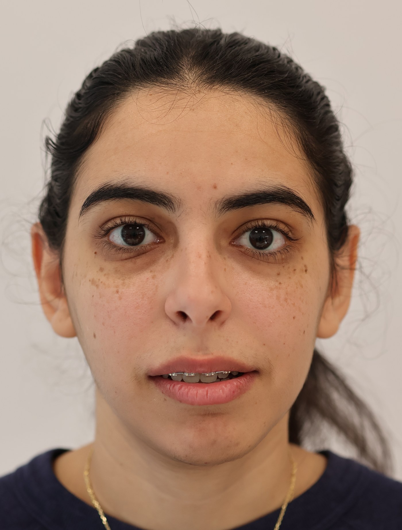
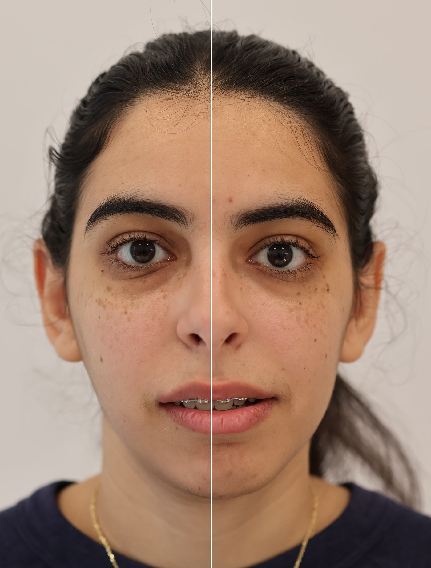
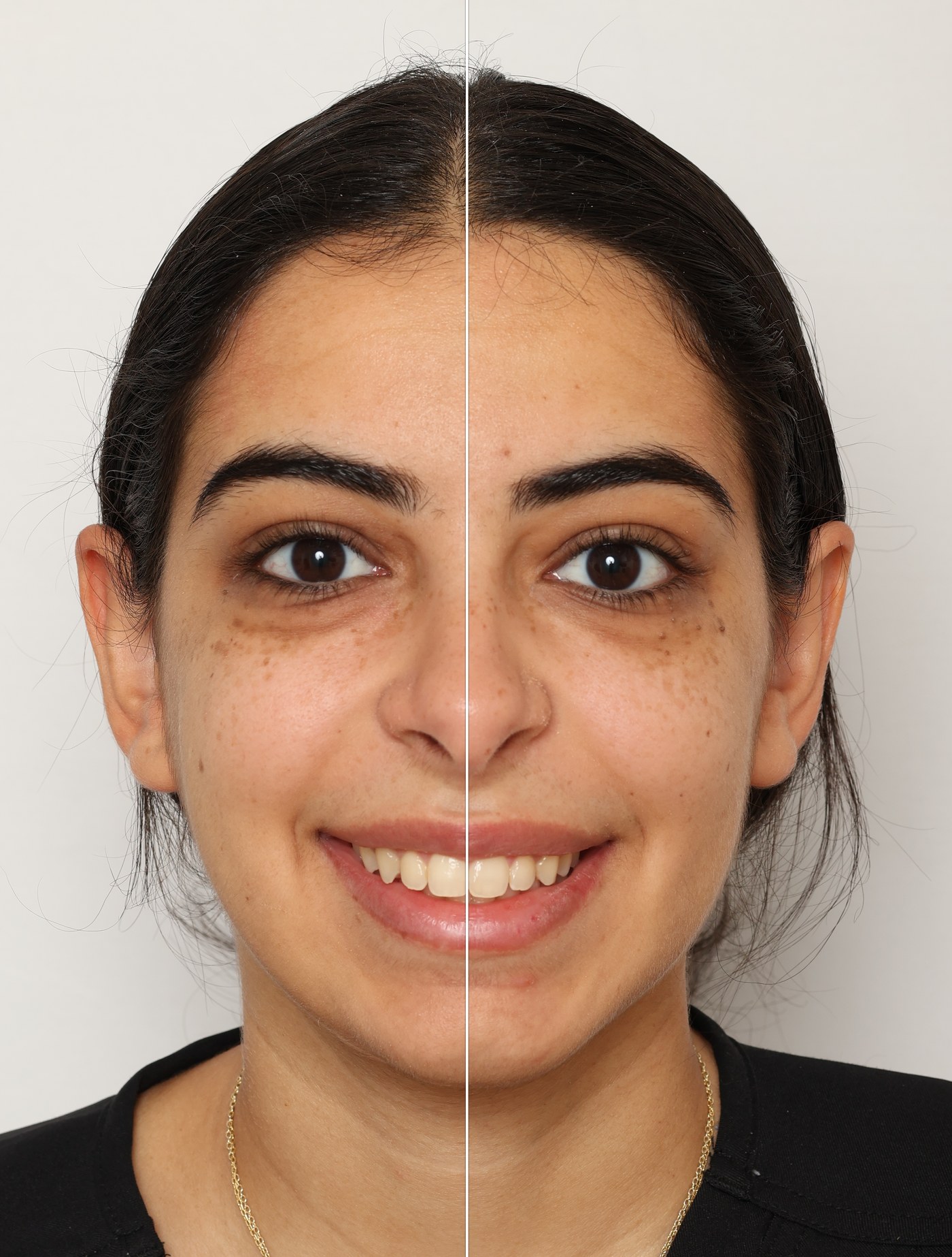
Individual results vary.
Centering the chin and the dental midlines
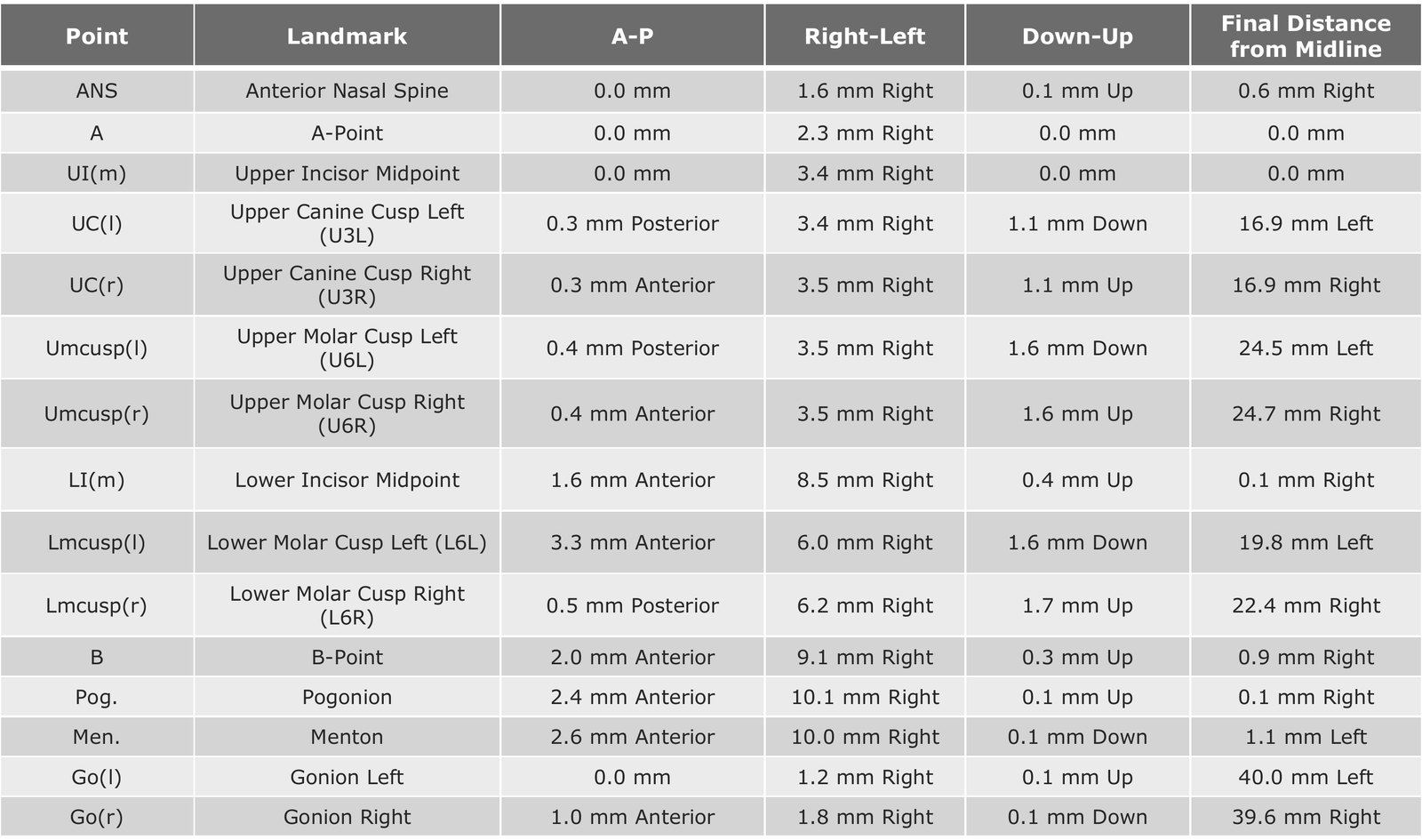
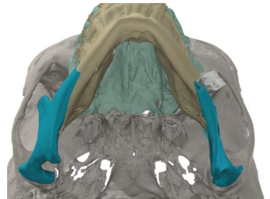
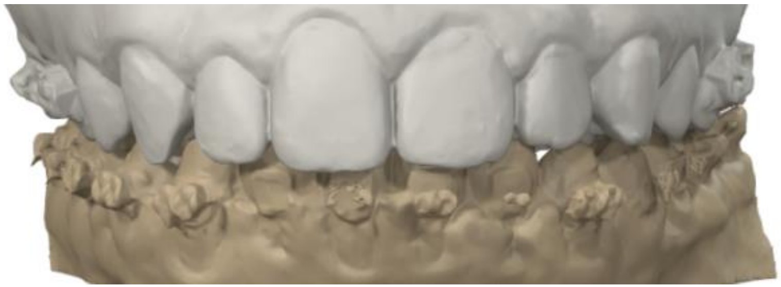
In the surgical plan, the chin (pogonion) was moved 10.1 mm toward the patient’s right, ending 0.1 mm from the facial midline. The lower incisor midline moved 8.5 mm to 0.1 mm from midline, and the upper incisor midline 3.4 mm to the midline. Forward movement was small (2.4 mm at the chin).
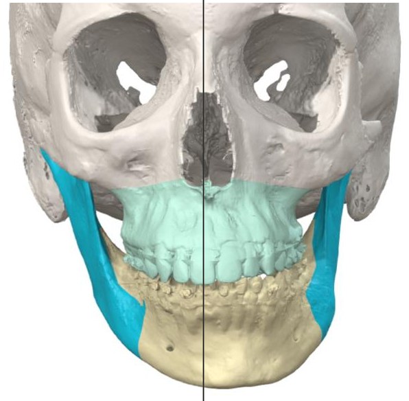 Before
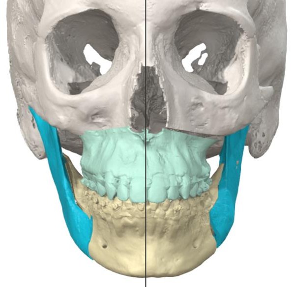 Planned
Diagnosis
Lower-face asymmetry with a tilted bite. Before surgery the chin (pogonion) sat 10.0 mm and the lower incisor midline 8.4 mm to the patient’s left of the facial midline, and the upper incisor midline 3.4 mm to the left. The bite was canted: the upper canines sat 47.1 mm (left) and 49.3 mm (right) below the eye-socket rim.
Measured on the 3D surgical plan before surgery.
Surgical plan
The movements were planned on a 3D model from the CT scan. In the surgical plan the upper jaw was rotated to level the occlusal plane: down 1.1 mm at the left canine and 1.6 mm at the left first molar, up by the same amounts on the right. The lower jaw was then brought to the centred upper jaw.
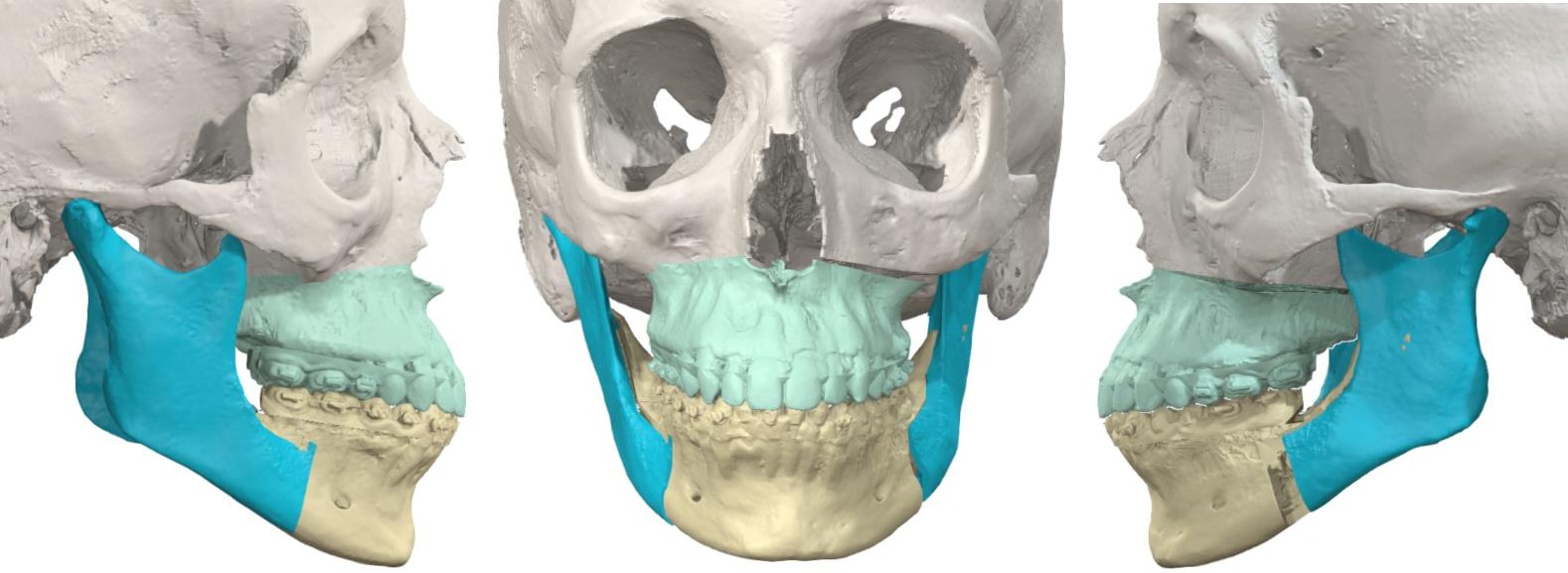
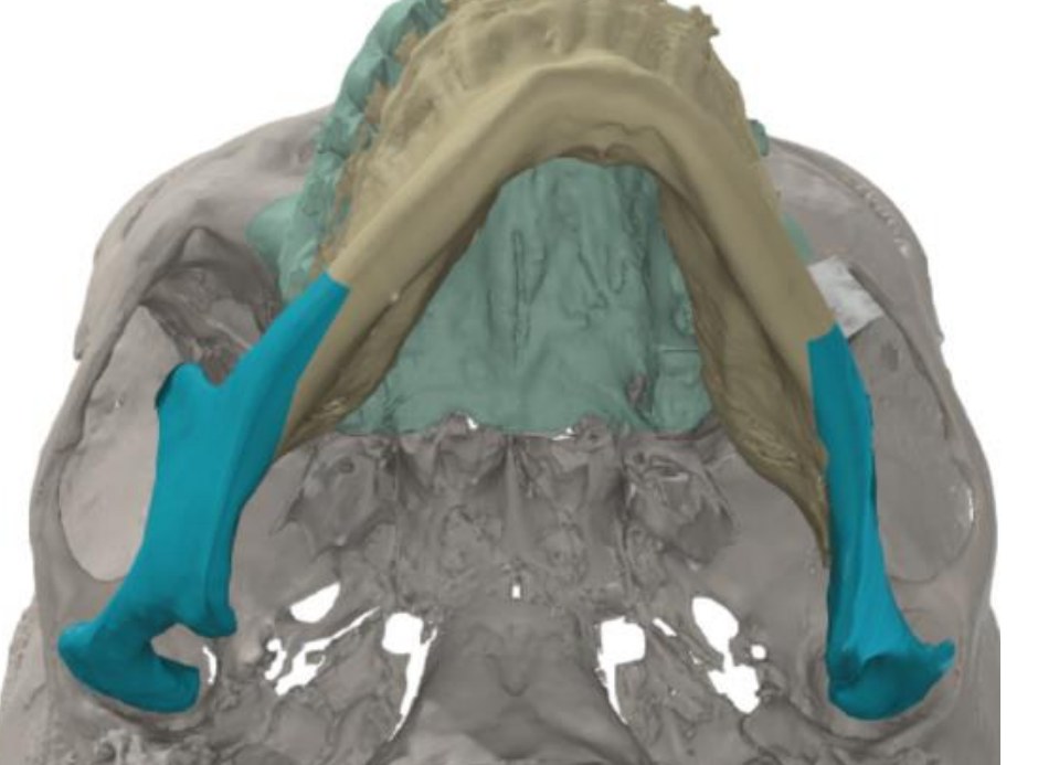 Before
 Planned
| Segment | Planned movement | Amount (from plan) |
|---|---|---|
| Chin (pogonion) | Right / forward | 10.1 mm / 2.4 mm |
| Mandible (lower incisor) | Right / forward | 8.5 mm / 1.6 mm |
| Maxilla (upper incisor) | Right | 3.4 mm |
| Maxilla (canines) | Cant correction | Left 1.1 mm down, right 1.1 mm up |
| Maxilla (first molars) | Cant correction | Left 1.6 mm down, right 1.6 mm up |
| SNA / SNB | Change | 0.0° / +1.2° |
Procedure
- Upper jaw rotated to level the occlusal plane and centre the upper midline (Le Fort I osteotomy)
- Lower jaw rotated toward the right and brought slightly forward (bilateral sagittal split osteotomy)
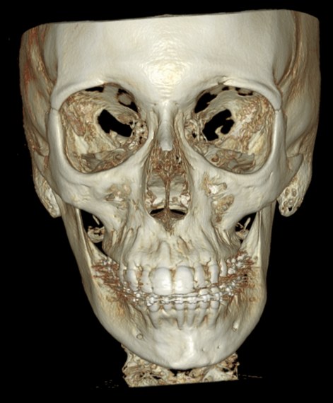
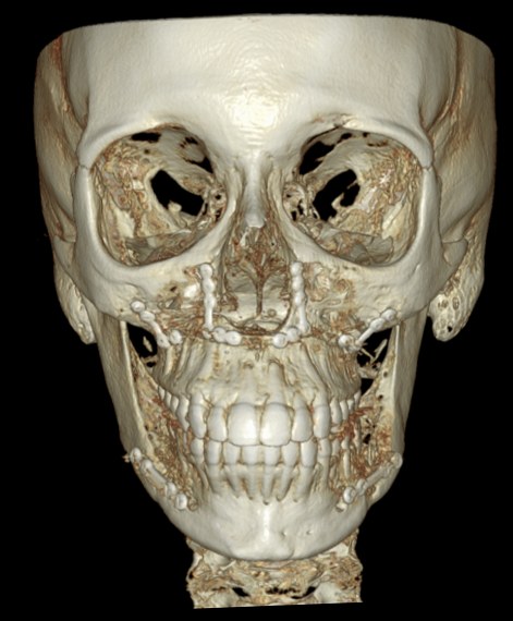
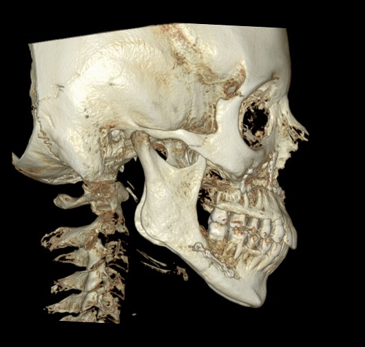
- Both jaws fixed with titanium plates and screws (visible on the post-operative CT)
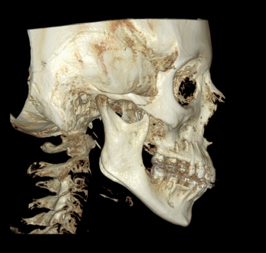
3D CT · before & after
Bone views from the CT scans before and after surgery, at the same angle and size. After surgery the chin and both dental midlines line up with the middle of the face, and the plates and screws are visible.
Individual results vary.
Where
Treated at Maxillo-Facial Surgery Institute, Irvine. Our Surgeons