Referring doctors: Send a referral (opens in a new tab)Our SurgeonsPatient resources17877 Von Karman Ave, Suite 370, Irvine(949) 771-2121
Home › Cases › Case 03
Case 03 · Jaw Surgery

Bimaxillary Surgery with Genioplasty: Front-to-Back Correction

A triple-jaw case planned to correct the front-to-back relationship of the jaws. The upper jaw was moved forward and up, the lower jaw was set back, and the chin was repositioned separately.

PlanningRecords & 3DFollow-up
Concern
Wanted to bite better
Procedure
Le Fort I, BSSO, genioplasty
Focus
Anteroposterior: maxilla forward, mandible back
Diagnosis
Skeletal Class III with anterior crossbite
Age range
20s
Follow-up shown
6 months after surgery
01

Front-to-back correction

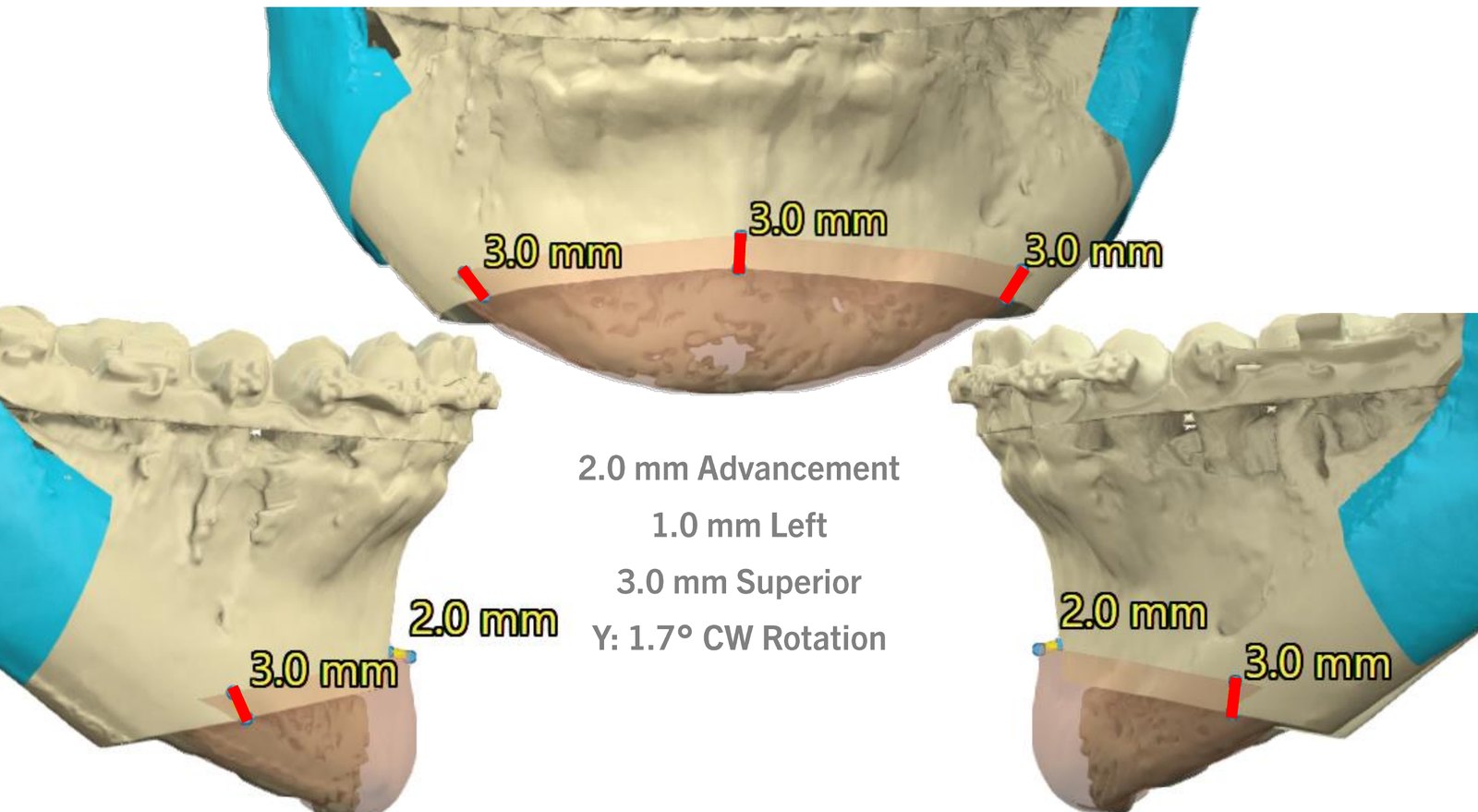
The upper jaw was planned 4.0 mm forward and 3.0 mm up. The lower jaw was set back about 5 mm at the lower incisor and B-point. The chin was then brought forward 2.0 mm and up 3.0 mm with a genioplasty.
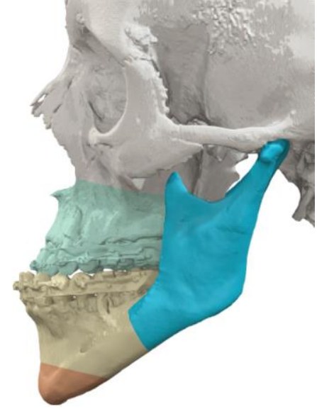
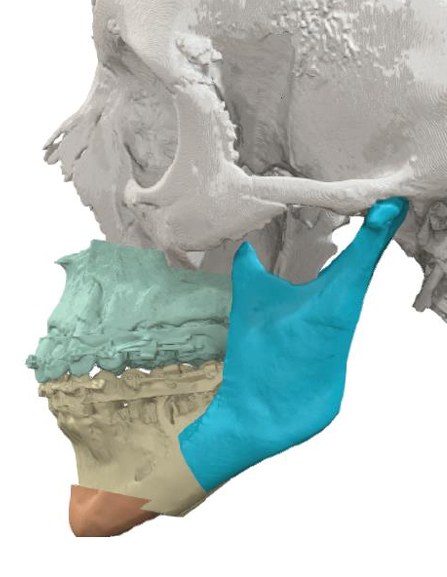
Maxilla
+4.0mm forward
and 3.0 mm up
Mandible
−4.7mm back
lower incisor; B-point 5.1 mm
Chin (genioplasty)
+2.0mm forward
3.0 mm up, 1.0 mm left
Lateral · Before
Lateral · BeforePre-operative position
Lateral · Planned
Lateral · PlannedFinal position
Genioplasty
GenioplastyPlanned chin movement
02

Diagnosis & concern

Concern

Her main concern was her bite. In her words, she wanted “to finally bite better.” Her lower front teeth closed in front of the upper front teeth, and her profile was concave.

Diagnosis

Severe skeletal Class III: the upper jaw sat back and the lower jaw forward, with the chin point set back. The bite was Class III with an anterior crossbite (lower front teeth 4 mm ahead of the upper). The upper dental midline sat 2 mm to the right, with a mild tilt of the upper jaw.

Recommended at consultation: Le Fort I osteotomy, bilateral sagittal split osteotomy and sliding genioplasty.

03

Surgical plan

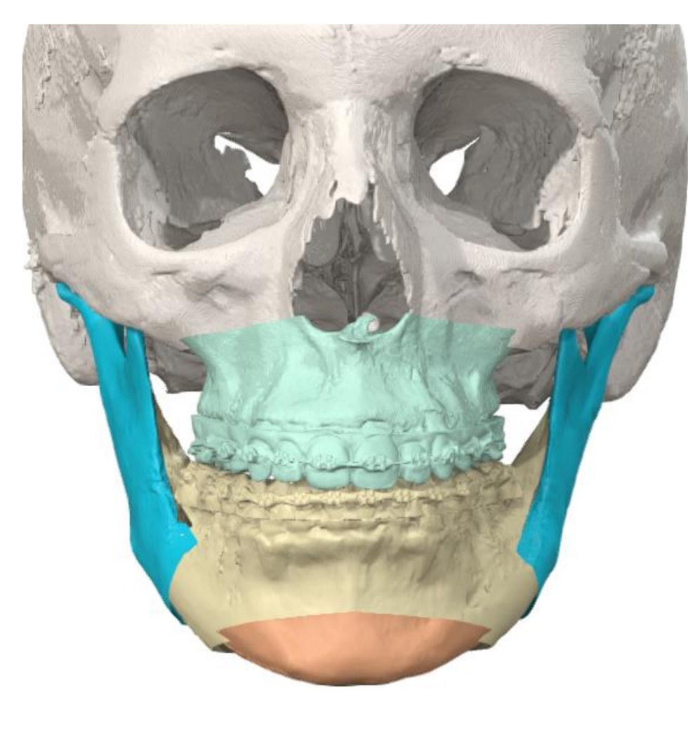
The movements were planned on a 3D model from the CT scan, upper jaw first, and transferred to surgery with splints, cutting guides and patient-specific plates.
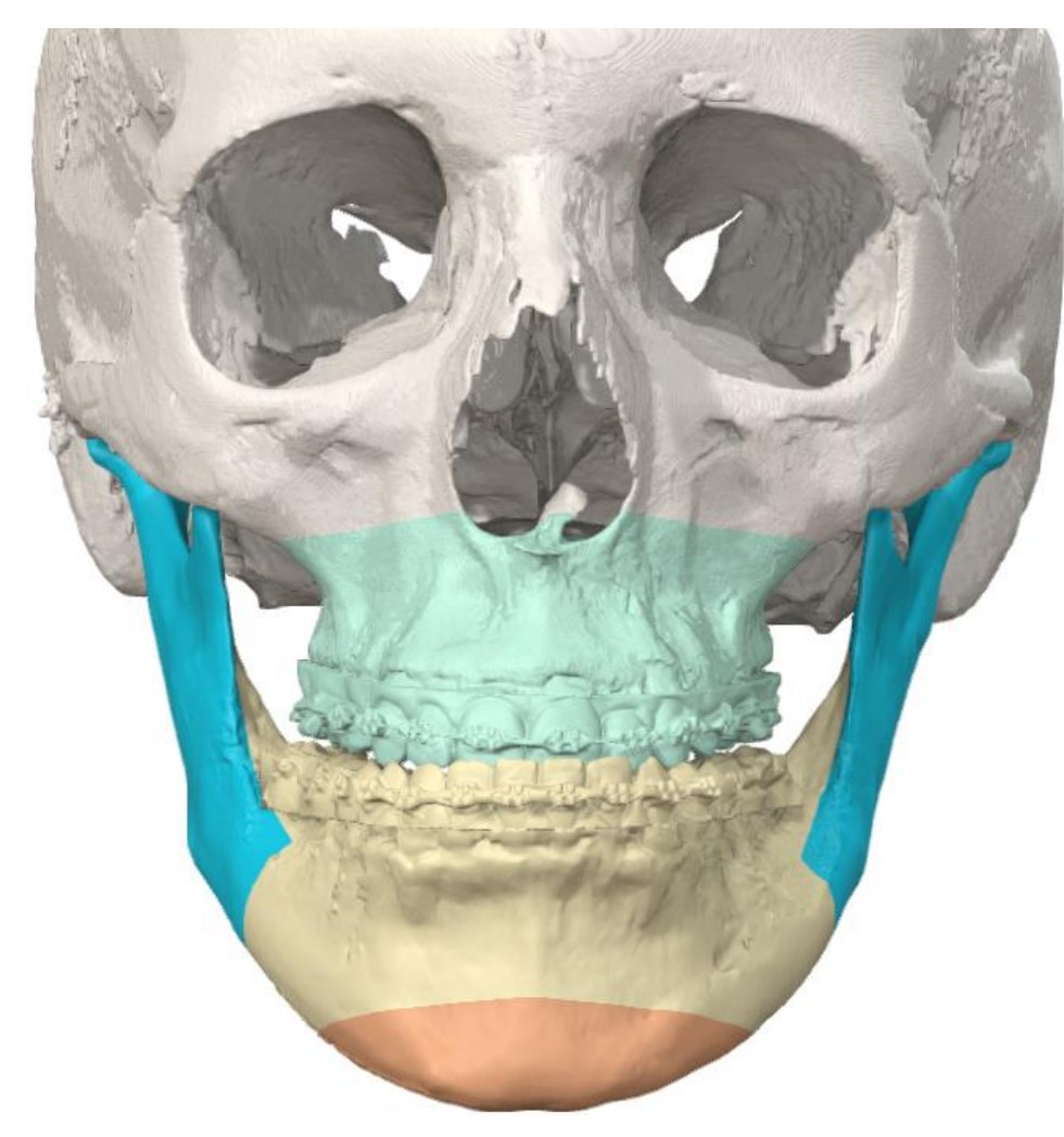
3D · Before · beforeBefore
3D · Before · plannedPlanned
3D · Before → plannedFrontal view; maxilla 4.0 mm forward, 3.0 mm up
SegmentPlanned movementAmount (from plan)
Maxilla (upper incisor)Forward / up4.0 mm / 3.0 mm
Mandible (lower incisor)Back / up4.7 mm / 3.2 mm
Mandible (B-point)Back5.1 mm
Chin (genioplasty)Forward / up / left2.0 / 3.0 / 1.0 mm
SNA / SNBChange+4.4° / −2.9°
Values as listed in the 3D surgical plan. Planned values; the operative result may differ slightly.
04

Procedure

  1. Upper jaw moved forward and up (Le Fort I osteotomy)
  2. Lower jaw set back (bilateral sagittal split osteotomy)
  3. Chin moved forward and up (sliding genioplasty)
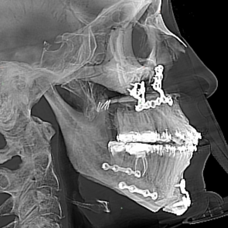
  4. Fixation with patient-specific plates (visible on the 6-month X-ray)
05

Records

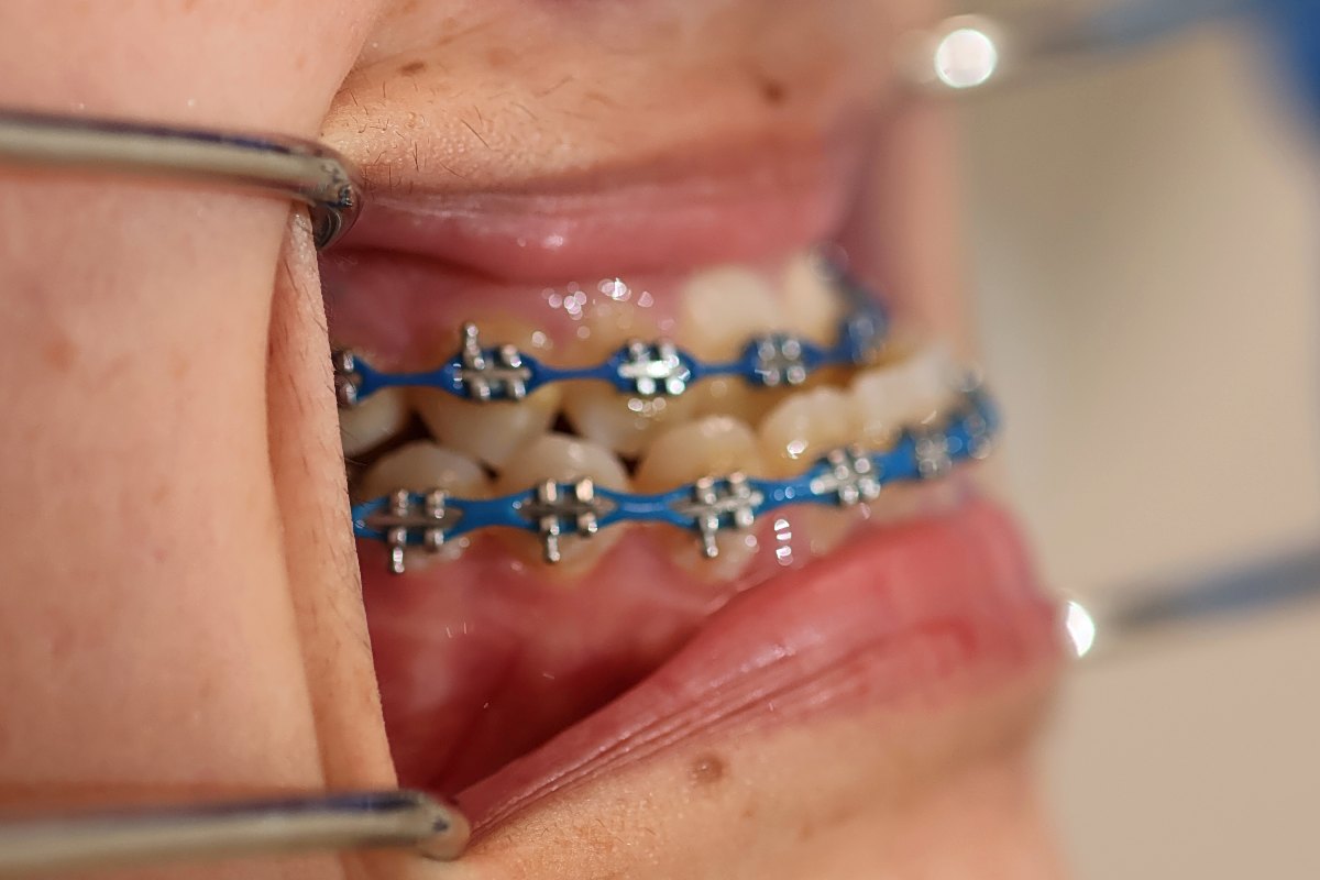
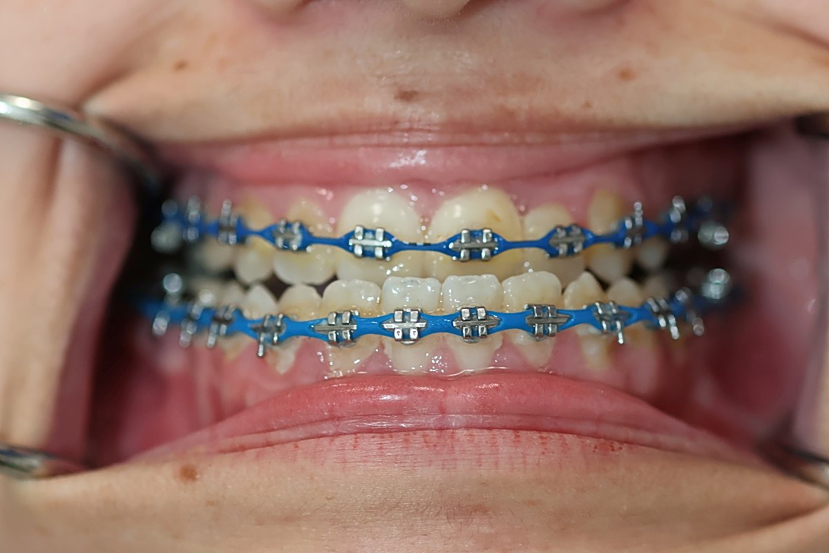
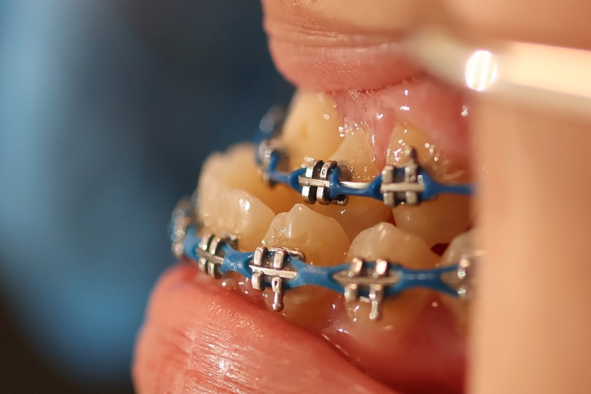
Bite photos · Before (pre-surgical orthodontics)
Bite photo before surgery, right side, with braces
Right sideBefore (pre-surgical orthodontics)
Bite photo before surgery, front, with braces
FrontBefore (pre-surgical orthodontics)
Bite photo before surgery, left side, with braces
Left sideBefore (pre-surgical orthodontics)
No post-operative bite photos are available.
View clinical records
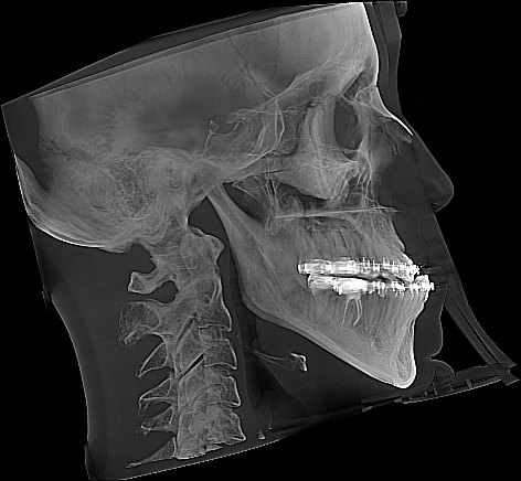
Lateral cephalometric X-ray before surgery, with braces
Ceph · BeforeShown as taken · see the 6-month X-ray in Follow-up
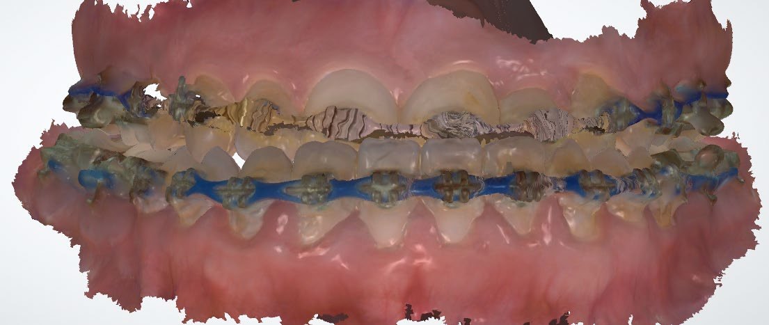
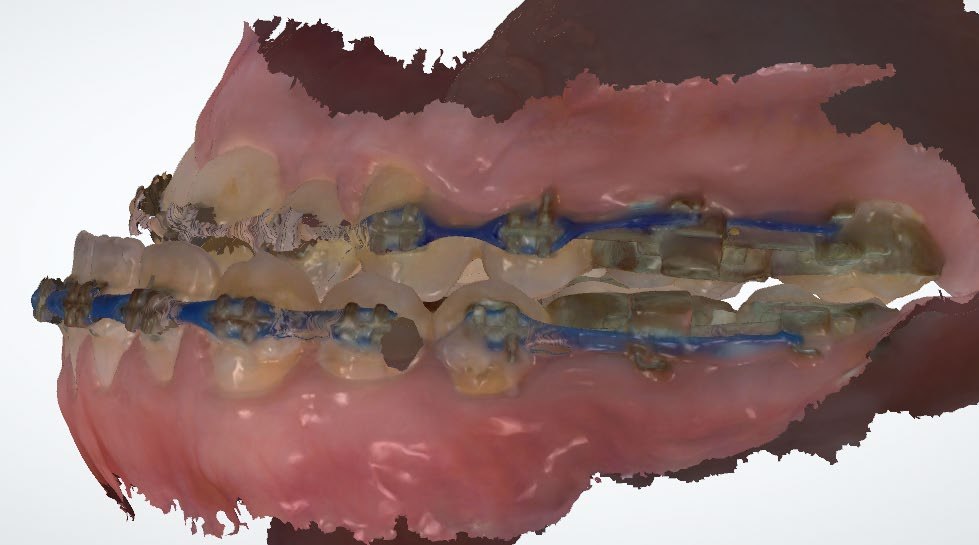
Intraoral scan before surgery, front view of the bite
Intraoral scan · Before · FrontFrom the consultation
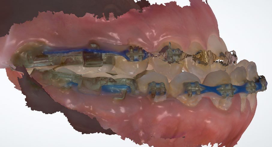
Intraoral scan before surgery, side view of the bite
Intraoral scan · Before · SideFrom the consultation
Intraoral scan before surgery, other side of the bite
Intraoral scan · Before · Other sideFrom the consultation
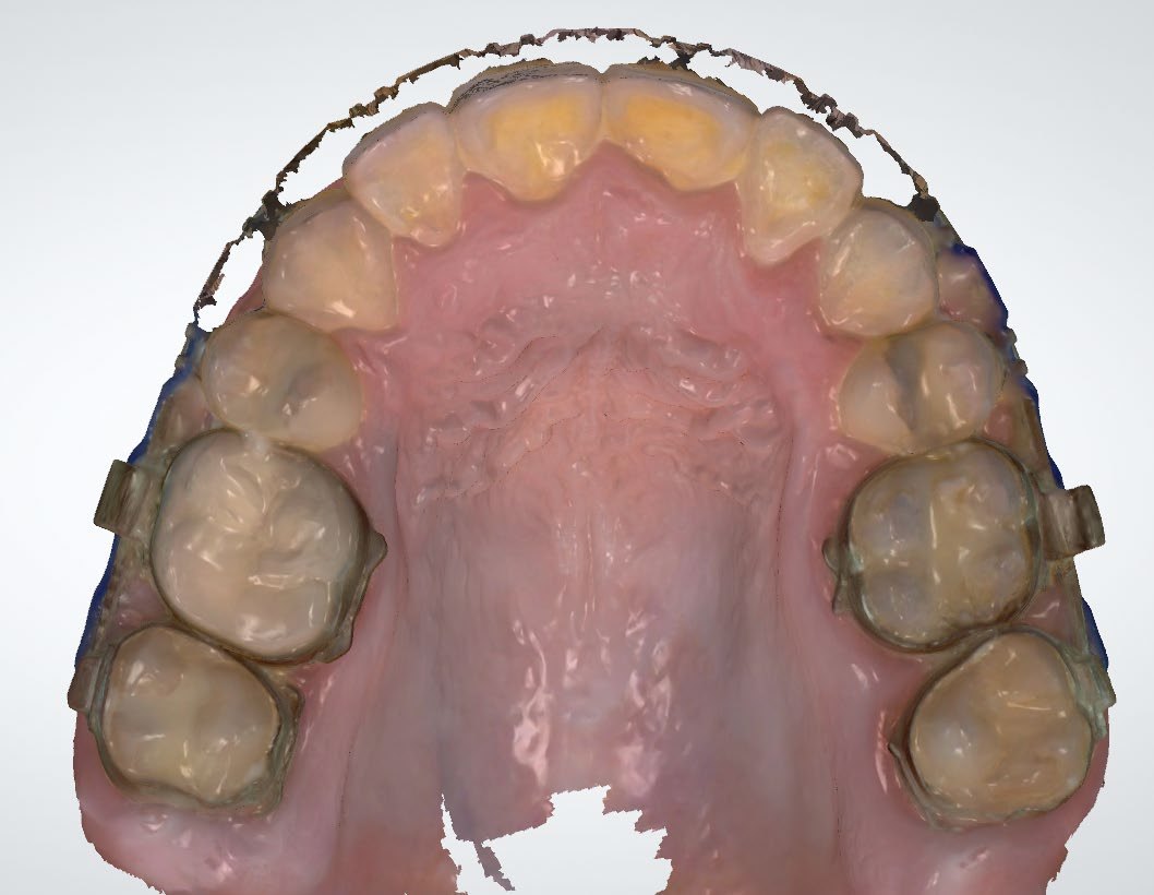
Intraoral scan before surgery, upper arch from below
Intraoral scan · Before · Upper archFrom the consultation
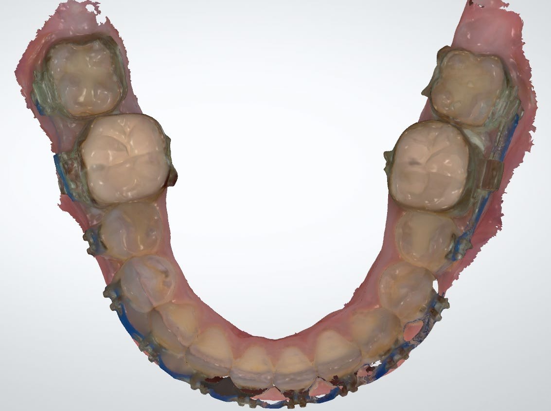
Intraoral scan before surgery, lower arch from above
Intraoral scan · Before · Lower archFrom the consultation
Intraoral scans shown as provided, without edits.
06

Follow-up: 6 months

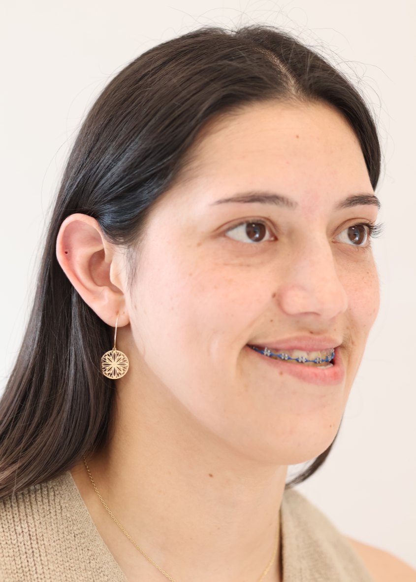
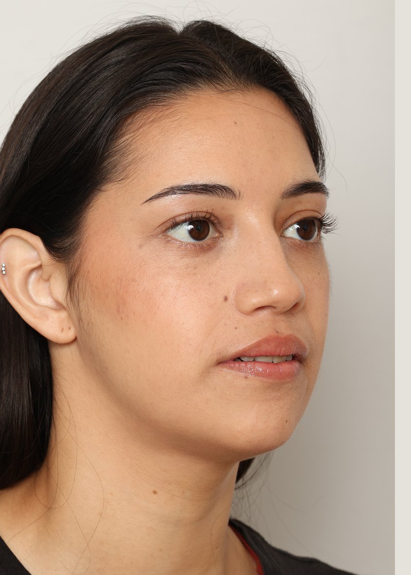
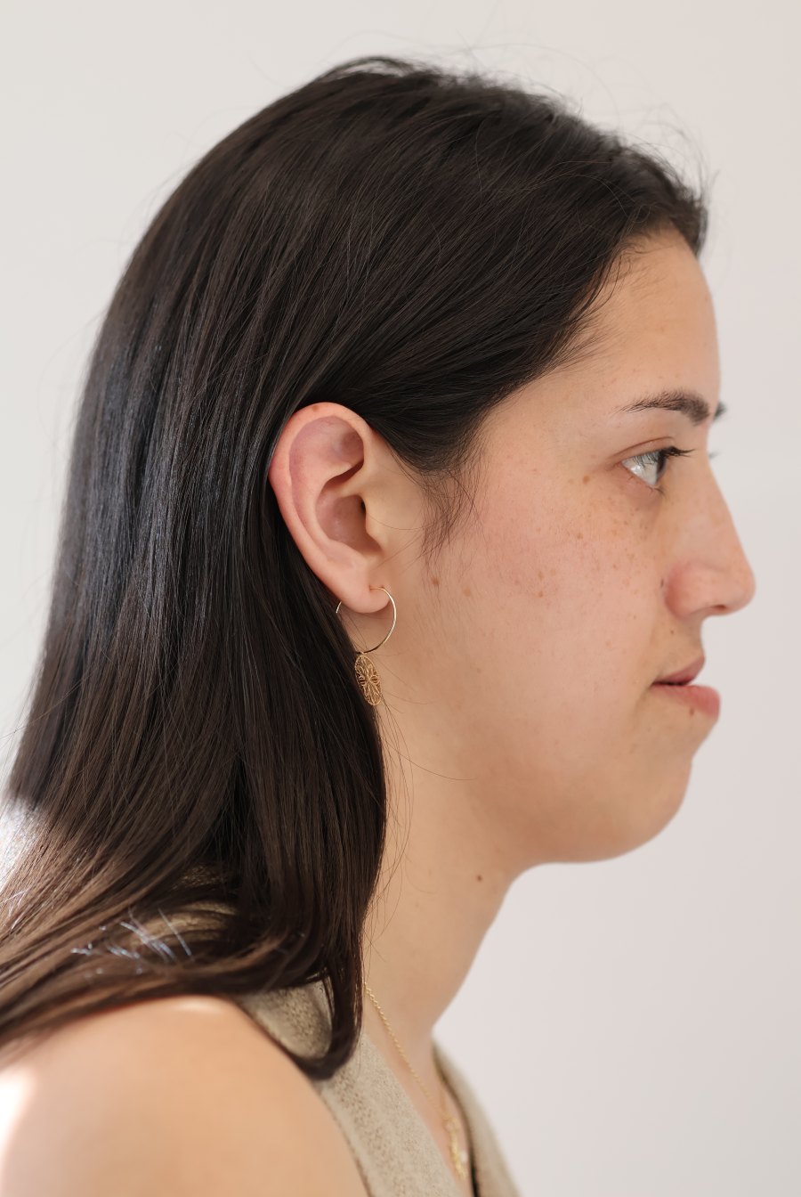
Photographs before surgery, during pre-surgical orthodontics (braces), and 6 months after surgery, with a lateral cephalometric X-ray.
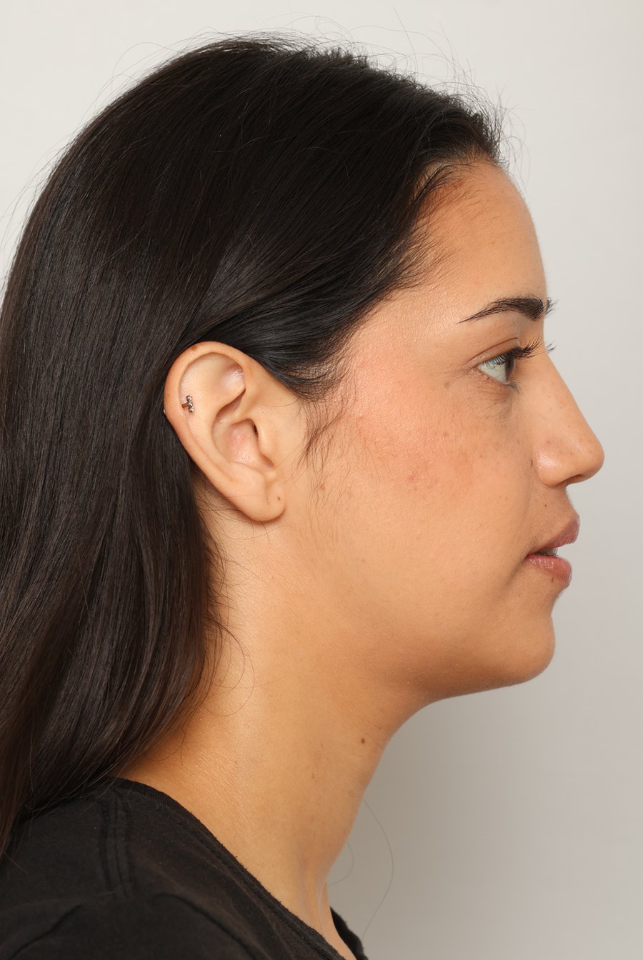
Right profile view, 6 months after surgery
Right profile view before surgery, with braces
BEFOREAFTER
‹ ›
Right profile · Before / After (6 months)Drag to compare. Before: pre-surgical orthodontics
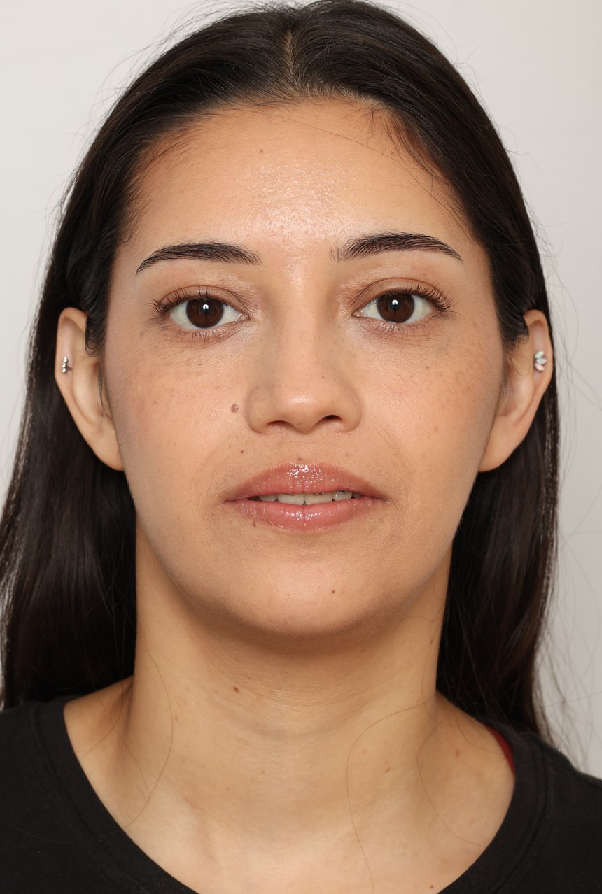
Front
Front view before surgery, with braces
BeforePre-surgical orthodontics
Front view, 6 months after surgery
After6 months
Three-quarter (right) · Before smiling, After at rest
Right three-quarter view before surgery, with braces
BeforePre-surgical orthodontics
Right three-quarter view, 6 months after surgery
After6 months
Profile (right)
Right profile view before surgery, with braces
BeforePre-surgical orthodontics
Right profile view, 6 months after surgery
After6 months
Cephalometric X-ray · Before / After (6 months)
Lateral cephalometric X-ray before surgery, with braces
Ceph · BeforeBefore surgery
Lateral cephalometric X-ray 6 months after surgery, with fixation plates
Ceph · After (6 months)Fixation plates visible
The two X-rays are not at the same scale or orientation.

Individual results vary.

07

Where

Treated at Maxillo-Facial Surgery Institute, Irvine. Our Surgeons

Plan images are cropped from the 3D surgical planning report; measurements are shown as planned. Individual results vary; this page is educational and not medical advice.
Call (949) 771-2121Request consultation