
Two-Piece Le Fort I and BSSO: Upper Jaw Widened and Moved Forward
A double-jaw case in which the upper jaw was divided into two segments and widened, then brought forward and up. The lower jaw was positioned to the new upper jaw. Planning and fixation were done with patient-specific guides and plates.
- Concern
- Wanted to bite better
- Procedure
- Two-piece Le Fort I with expansion; BSSO
- Focus
- Transverse: maxillary widening
- Diagnosis
- Skeletal Class III, narrow upper jaw, crossbite, open bite
- Age range
- 30s


Widening the upper jaw
The maxilla was planned in two segments, divided near the midline of the palate. The segments were opened 9.7 mm at the back, widening the arch across the first molars from 46.2 to 51.1 mm.
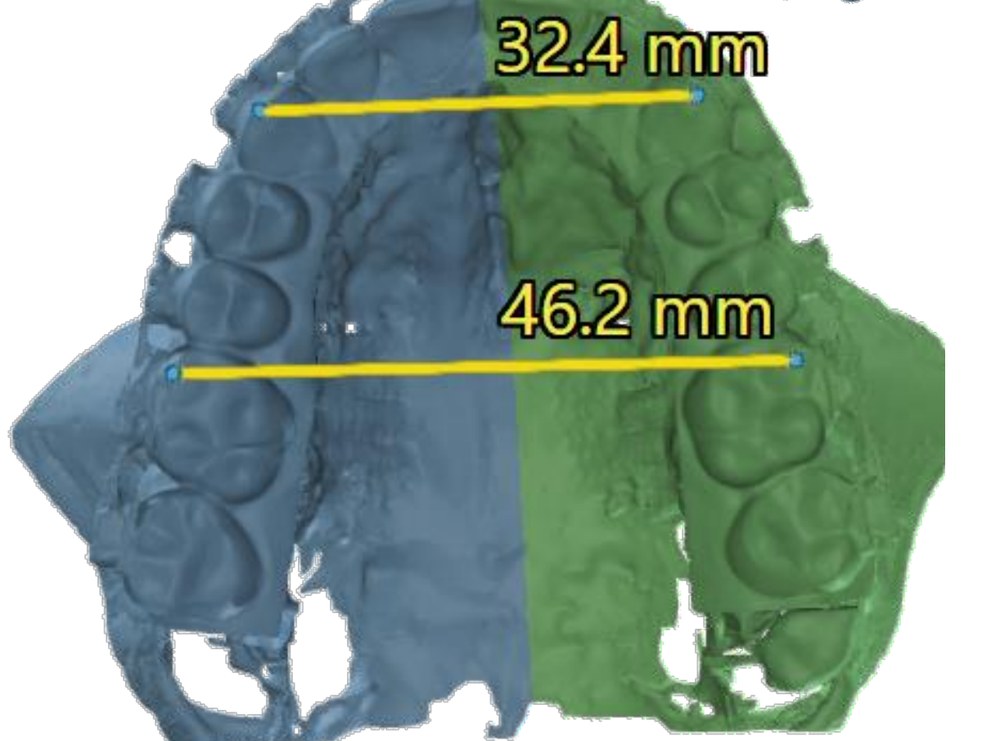 Before
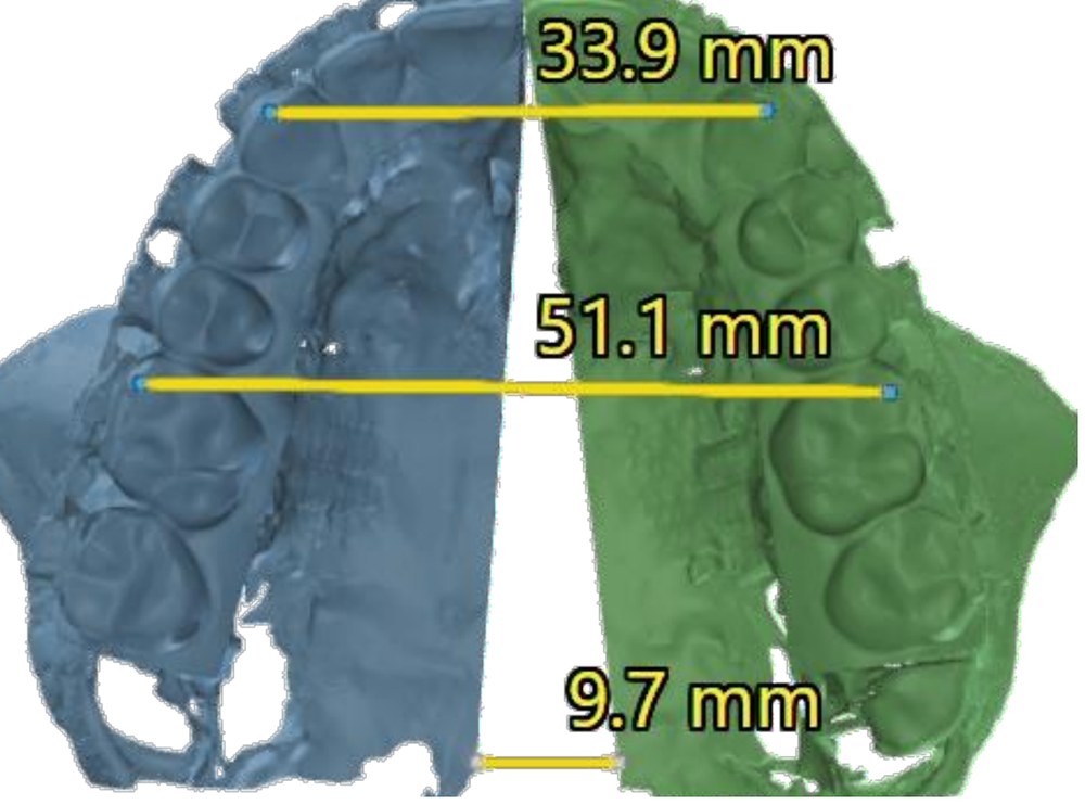 Planned
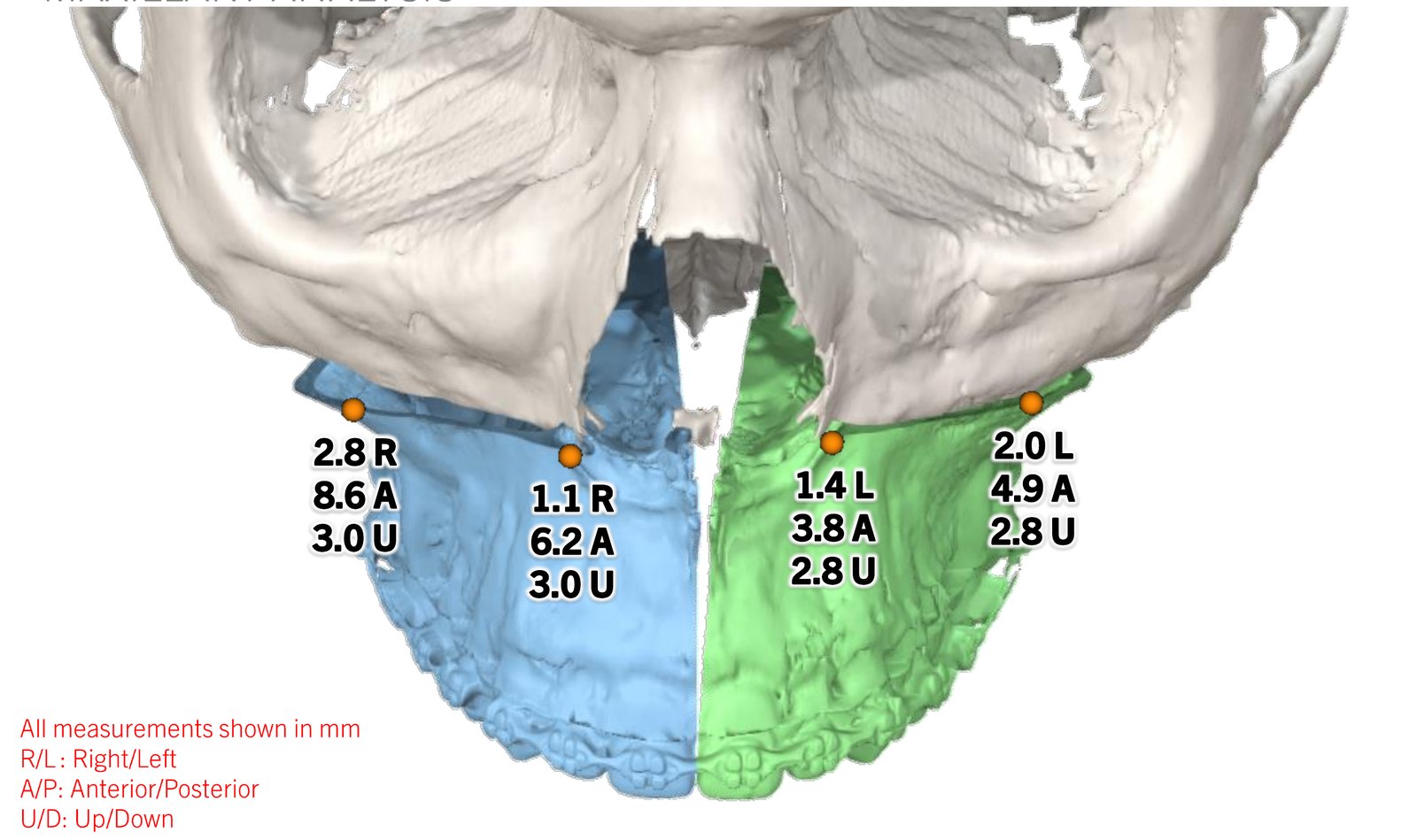
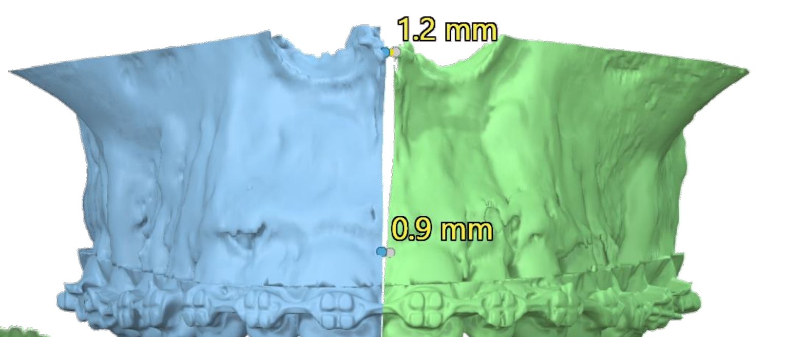
Diagnosis & concern
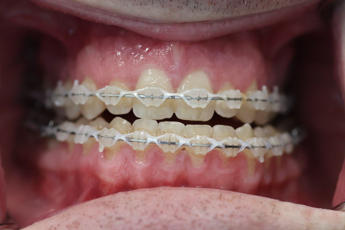
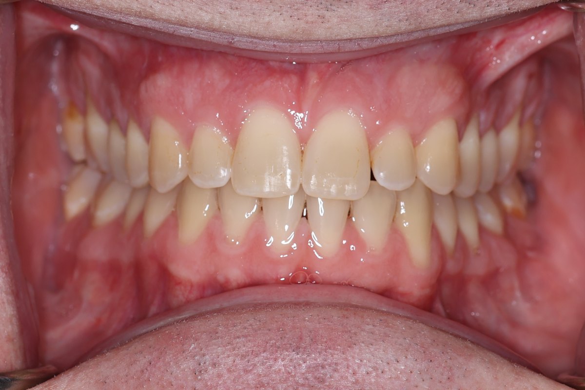
His main concern was his bite. In his words, he wanted “to finally bite better.” His front teeth did not overlap when he closed, and his profile was concave.
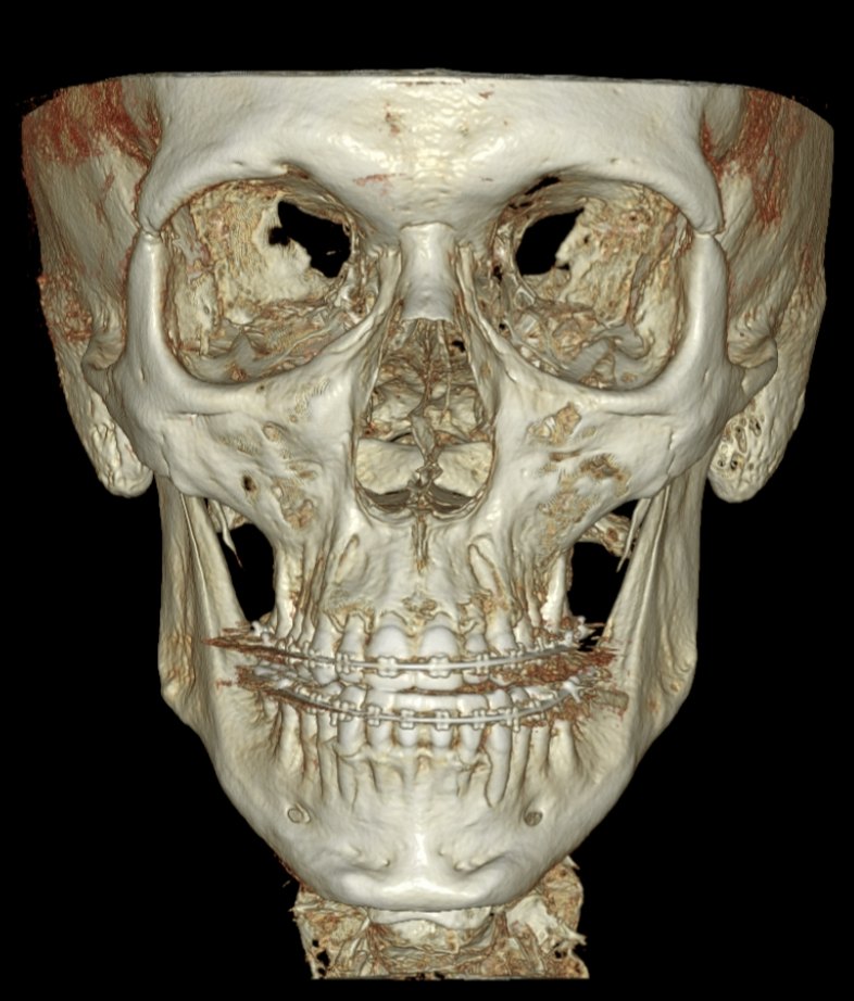
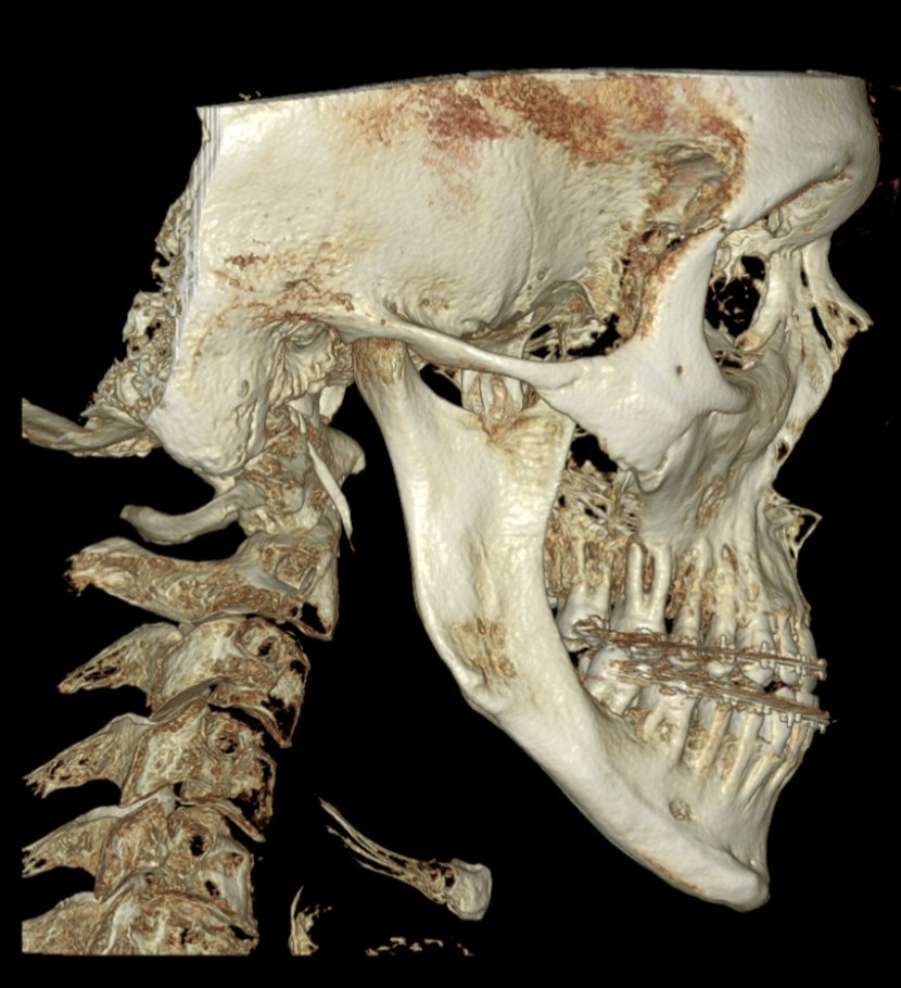
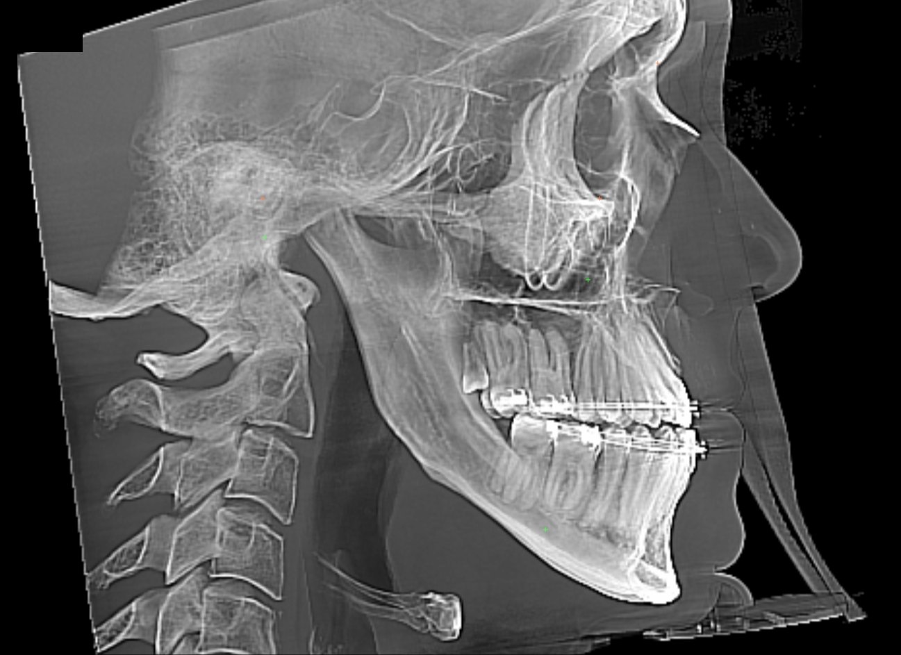
Skeletal Class III: the lower jaw sat forward of the upper jaw, with the chin slightly to the left. The upper jaw was narrow (about 5 mm too narrow across the back teeth), with a crossbite on the left side. The bite was Class III with an anterior open bite.
Surgical plan
The movements were planned on a 3D model from the CT scan, upper jaw first, and transferred to surgery with a palatal splint, cutting guides and patient-specific titanium plates for each maxillary segment.
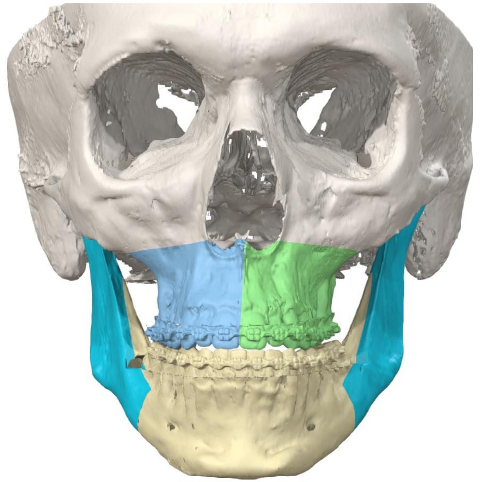 Before
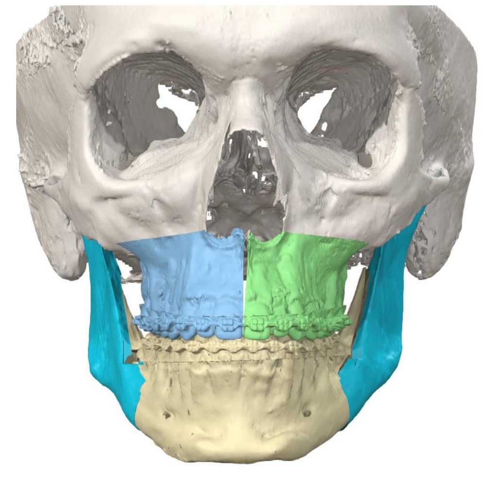 Planned
| Segment | Planned movement | Amount (from plan) |
|---|---|---|
| Maxilla (upper incisor) | Forward / up | 4.0 mm / 3.0 mm |
| Maxilla (molar width) | Widened, two segments | 46.2 → 51.1 mm |
| Mandible (lower incisor) | Back / up | 0.3 mm / 5.2 mm |
| Chin point (pogonion), moving with the lower jaw · no chin surgery | Forward / up | 2.1 mm / 5.1 mm |
| SNA / SNB | Change | +3.5° / +0.9° |
Procedure
- Upper jaw divided into two segments, widened, and moved forward and up (two-piece Le Fort I osteotomy)
- Lower jaw repositioned to the new upper jaw (bilateral sagittal split osteotomy)
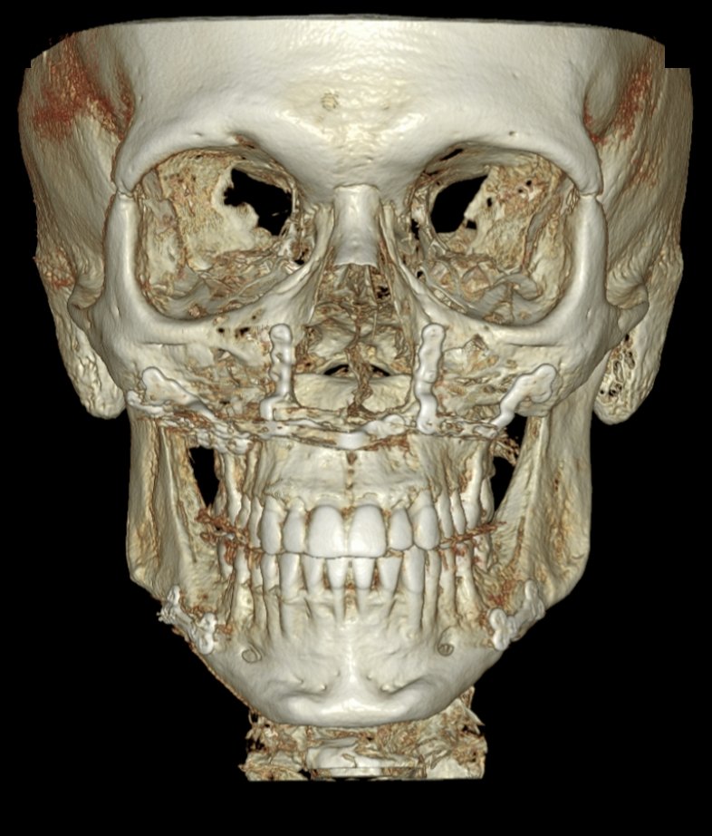
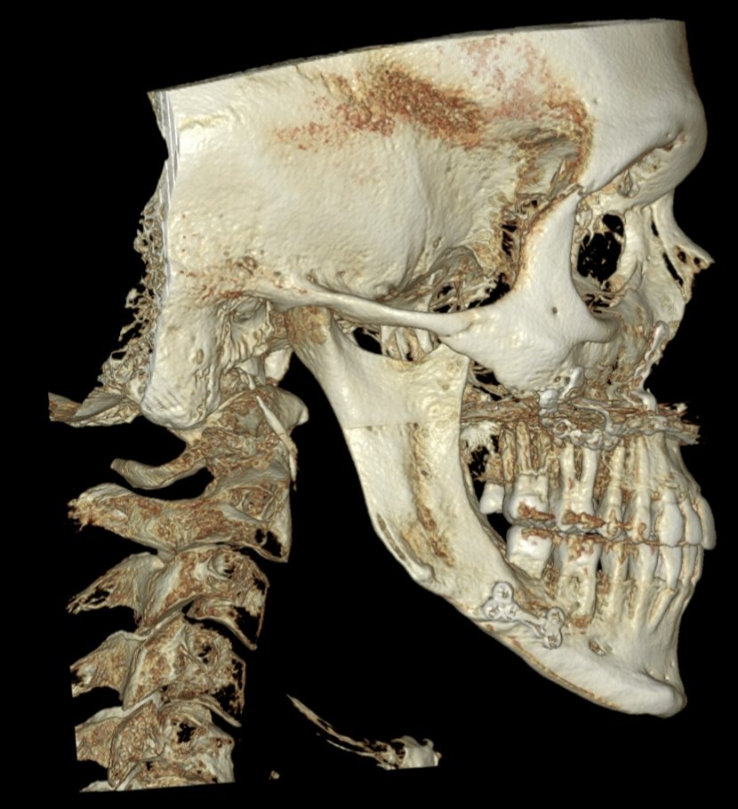
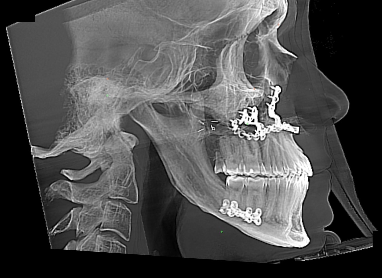
- Fixation with patient-specific titanium plates; palatal splint to hold the width. The plates are visible on the post-operative CT
3D CT · Before & after
3D CT before surgery and after surgery, shown at the same angle and scale. The fixation plates on the upper and lower jaw are visible after surgery.
Where
Treated at Maxillo-Facial Surgery Institute, Irvine. Our Surgeons