
Mandibular Midline Split with Segmental Maxillary Widening
A patient with an underbite and difficulty chewing. The lower arch was too wide for the upper arch. The key step was segmental surgery of the lower jaw: the mandible was split at the midline and narrowed from 45.8 to 43.4 mm across the molars. This reduced how far the upper jaw had to be widened, which was done in segments in the same operation. No genioplasty (chin surgery) was performed.
- Concern
- Underbite, difficulty chewing
- Procedure
- Mandibular midline osteotomy with BSSO; multi-piece Le Fort I with expansion; no genioplasty
- Focus
- Mandibular midline split (narrowing 45.8 → 43.4 mm), with segmental maxillary widening
- Diagnosis
- Skeletal Class III, narrow upper arch
- Age range
- 20s
- Follow-up shown
- 8 weeks after surgery
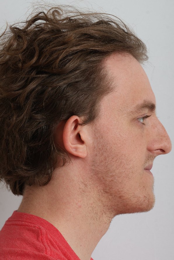
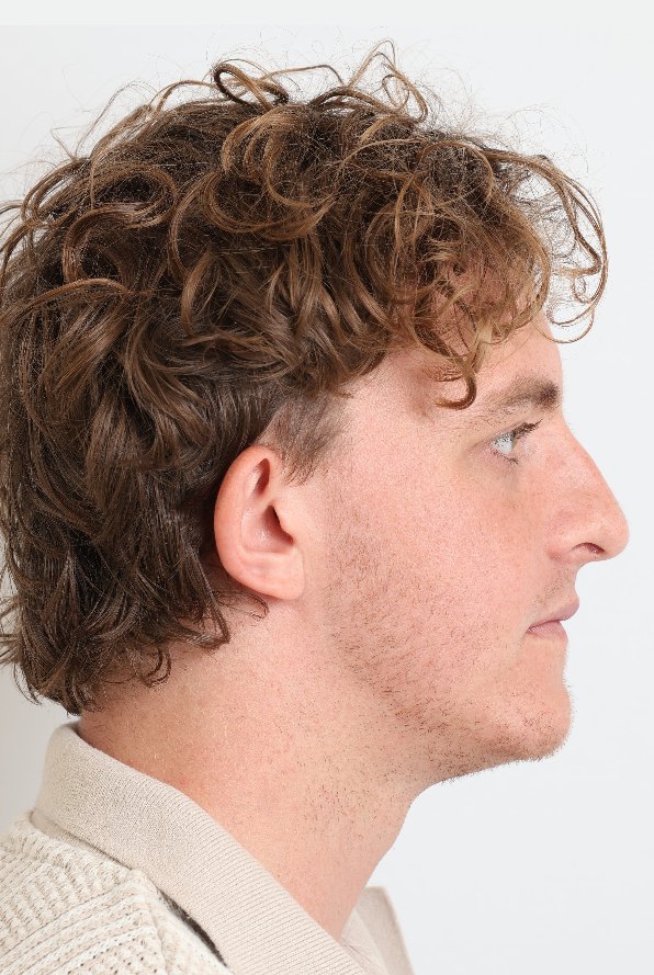
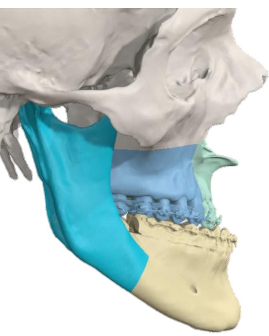
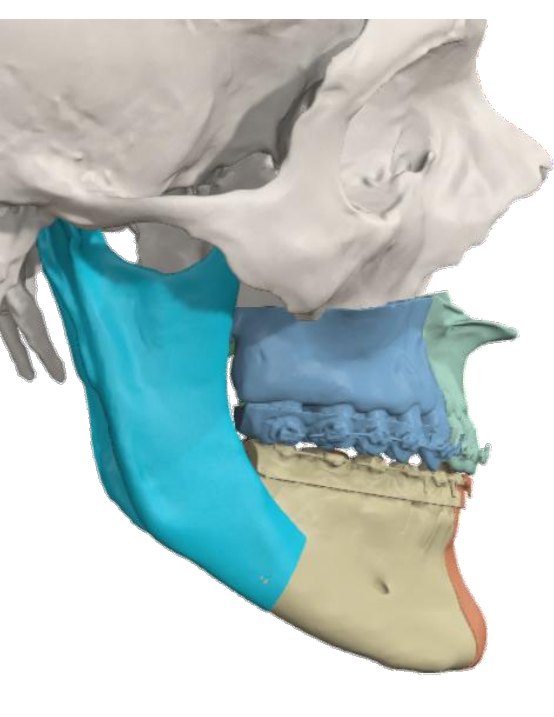
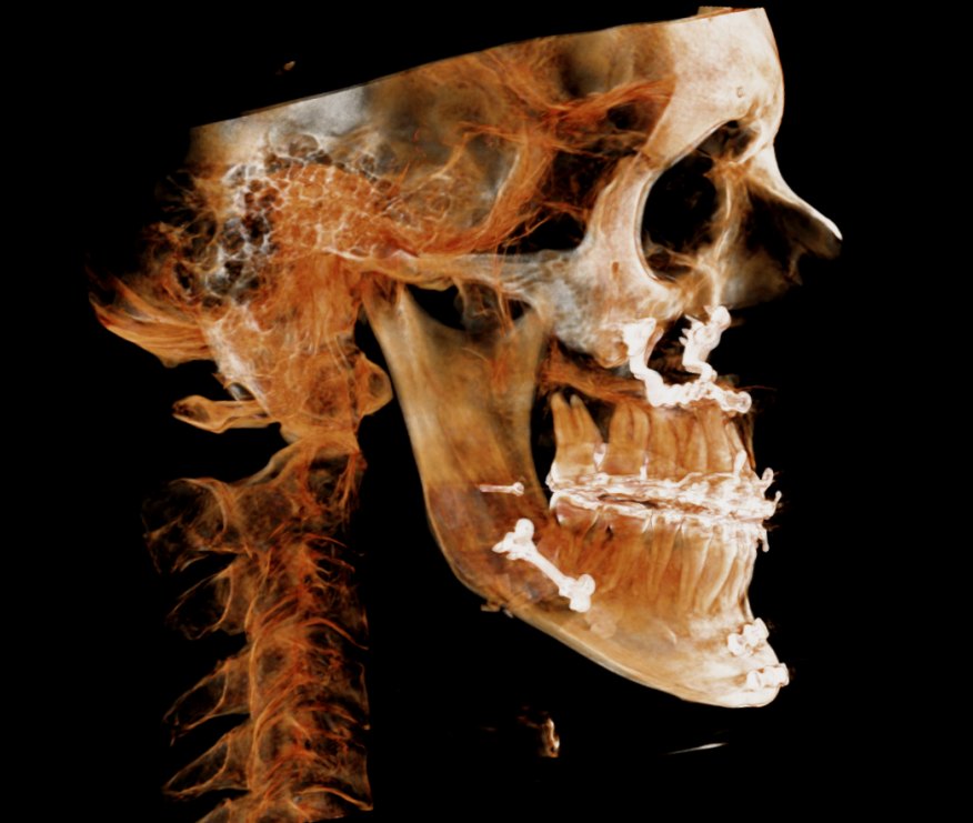
Splitting the lower jaw at the midline
The mandible was divided at the midline (midline mandibular osteotomy) and the two halves were brought together, narrowing the lower arch from 45.8 to 43.4 mm across the first molars. Narrowing the lower jaw reduced the widening needed in the upper jaw: 9.4 mm of opening between the posterior maxillary segments. The upper jaw was then widened in segments from 49.5 to 54.7 mm.
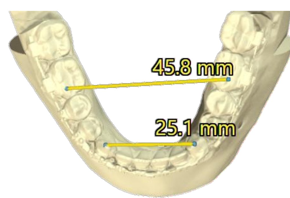 Before
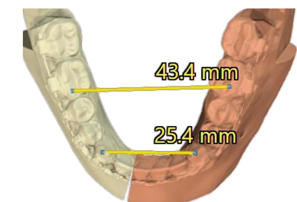 Planned
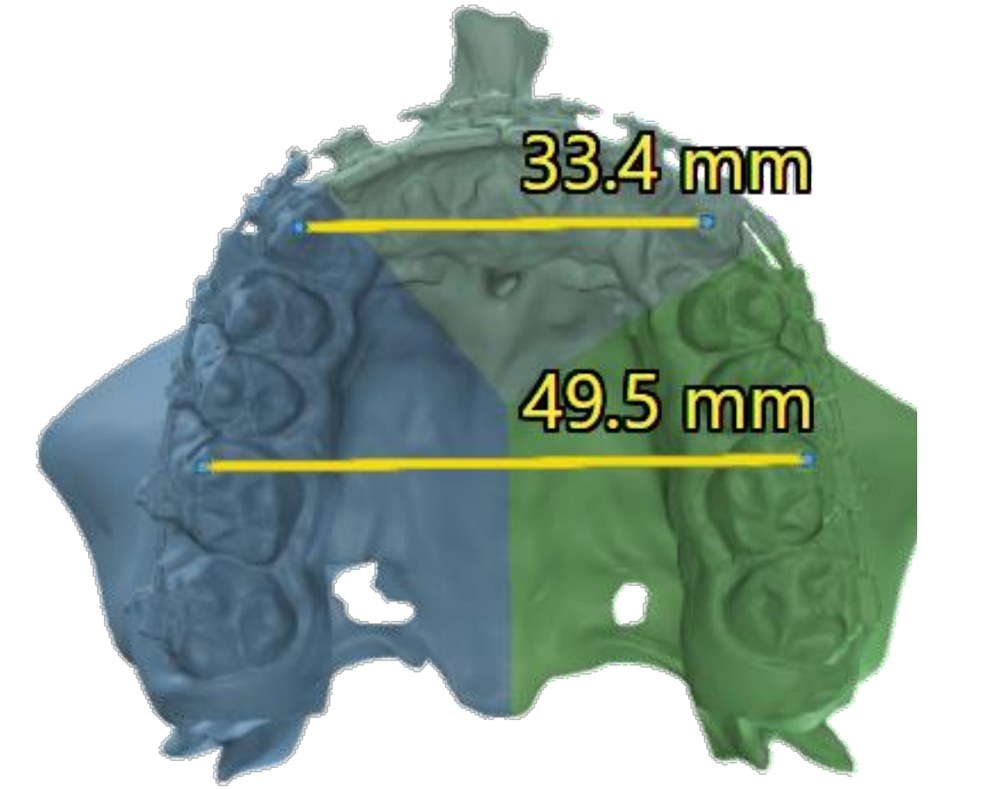 Before
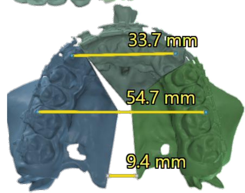 Planned
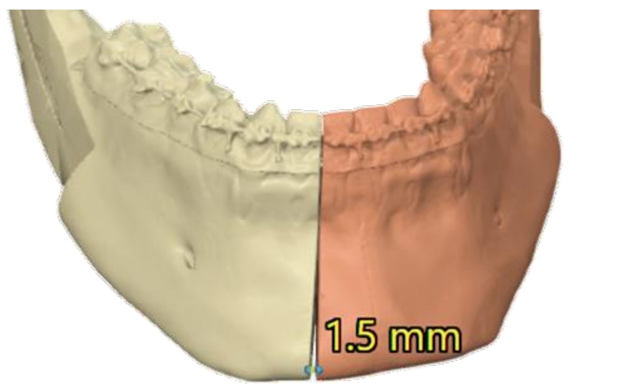
Diagnosis & concern
Underbite and difficulty chewing.
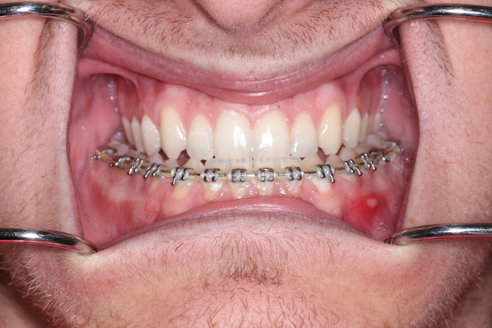
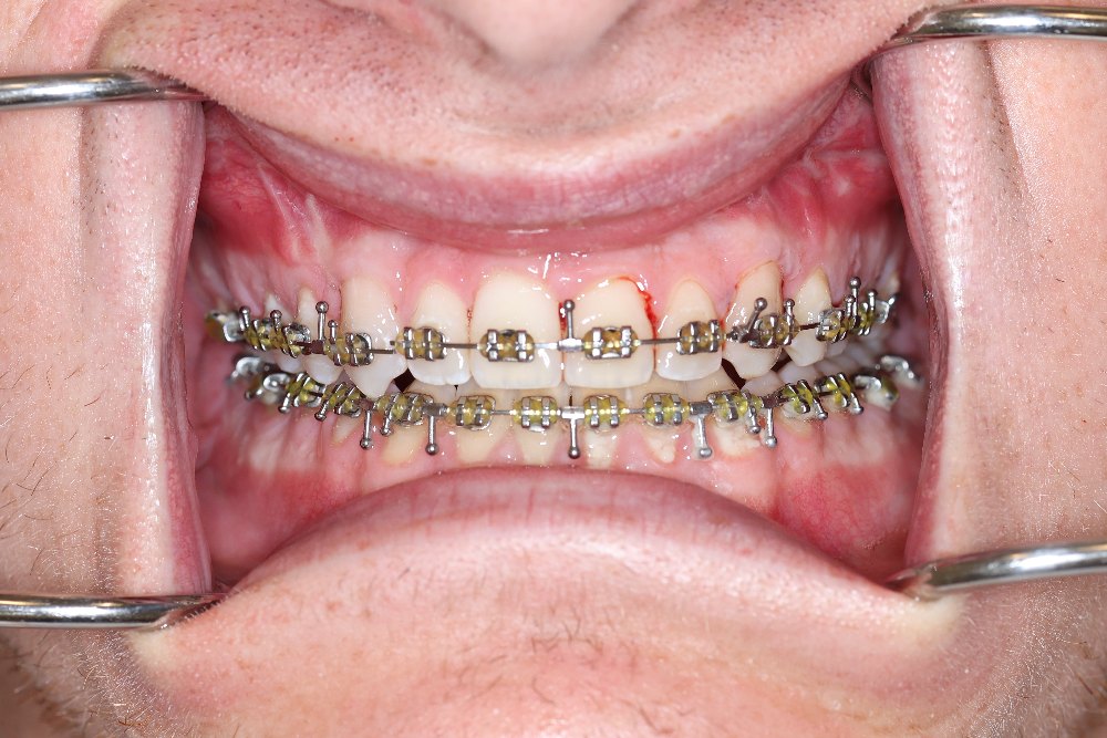
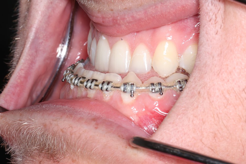
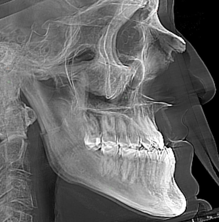
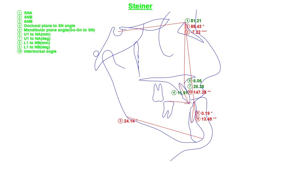
Skeletal Class III: the lower jaw and chin sat forward of the upper jaw (ANB −7.2° on the pre-operative X-ray), with a Class III bite and an anterior crossbite. There was also a transverse discrepancy: the upper arch was narrower than the lower arch. The chin and the dental midlines sat slightly to the left. Treatment addressed the bite and jaw relationship; there were no airway concerns.
Surgical plan
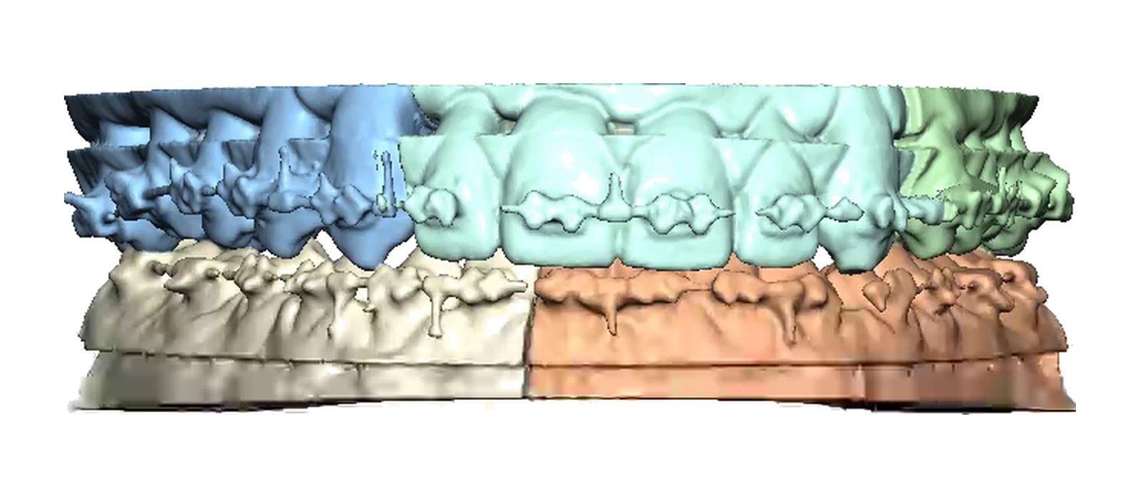
The movements were planned on a 3D model from the CT scan and transferred to surgery with splints. The upper jaw was planned first.
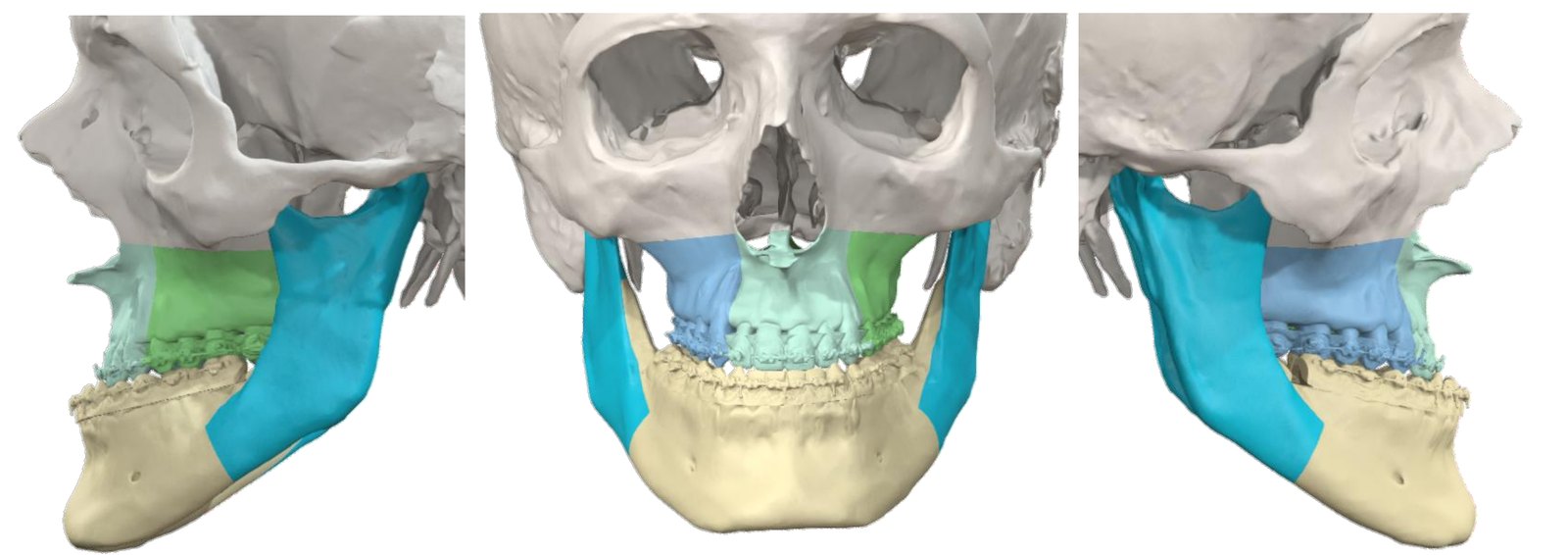
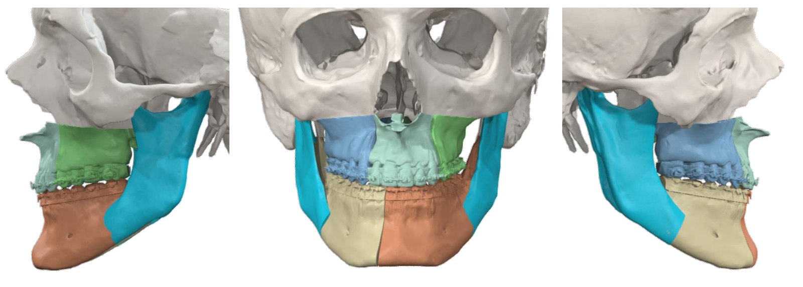
| Segment | Planned movement | Amount (from plan) |
|---|---|---|
| Mandible (molar width) | Split at midline, narrowed | 45.8 → 43.4 mm |
| Mandible (lower incisor) | Back / up | 3.4 mm / 4.1 mm |
| Maxilla (molar width) | Widened, segmental | 49.5 → 54.7 mm |
| Maxilla (upper incisor) | Forward / up | 7.0 mm / 2.0 mm |
| Chin | No genioplasty | None |
| SNA / SNB | Change | +5.1° / −1.4° |
Procedure
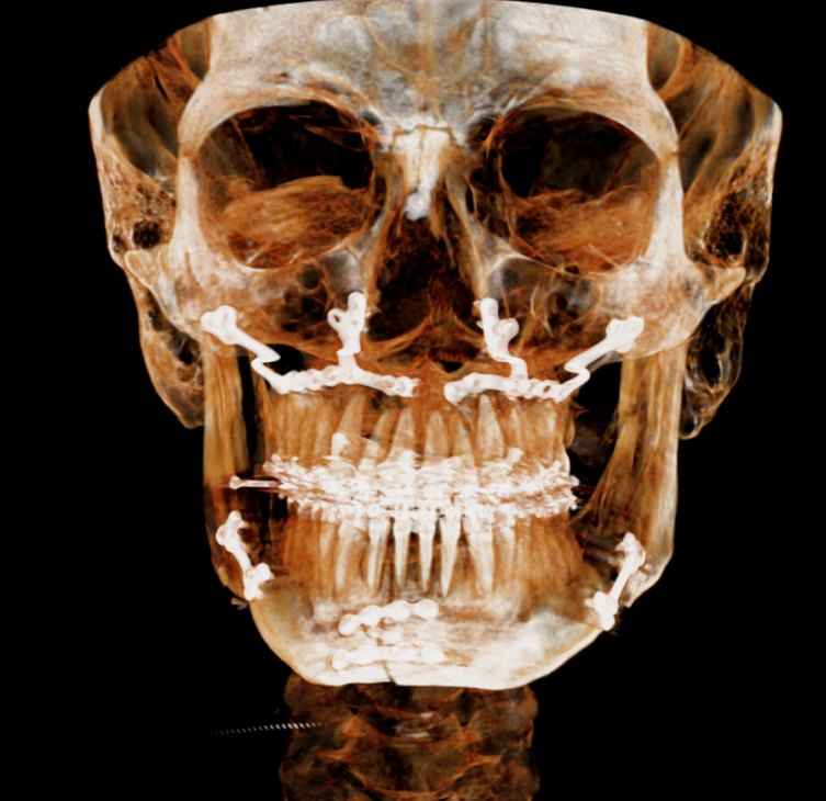
- Upper jaw repositioned and widened in segments (multi-piece Le Fort I osteotomy)
- Lower jaw divided at the midline and narrowed slightly (mandibular midline osteotomy)
- Lower jaw repositioned (bilateral sagittal split osteotomy)
- No genioplasty: the chin was not operated on
Records
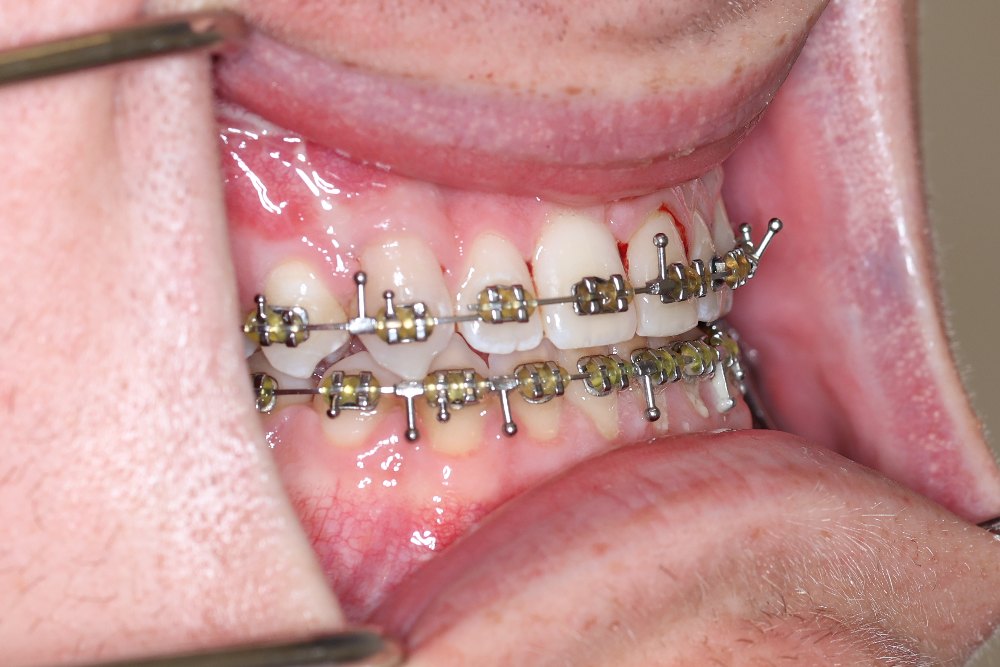
View clinical records
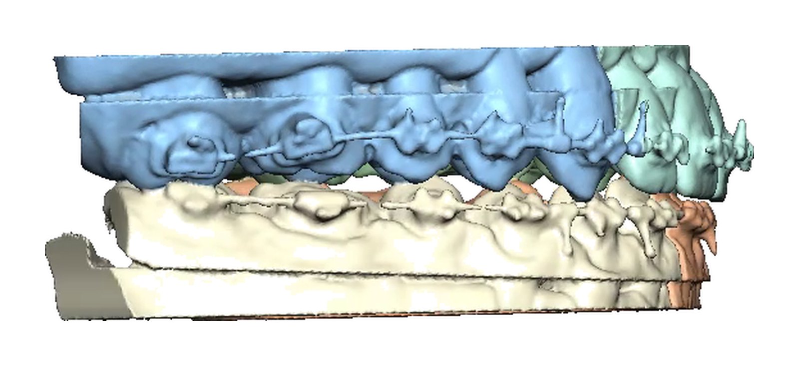
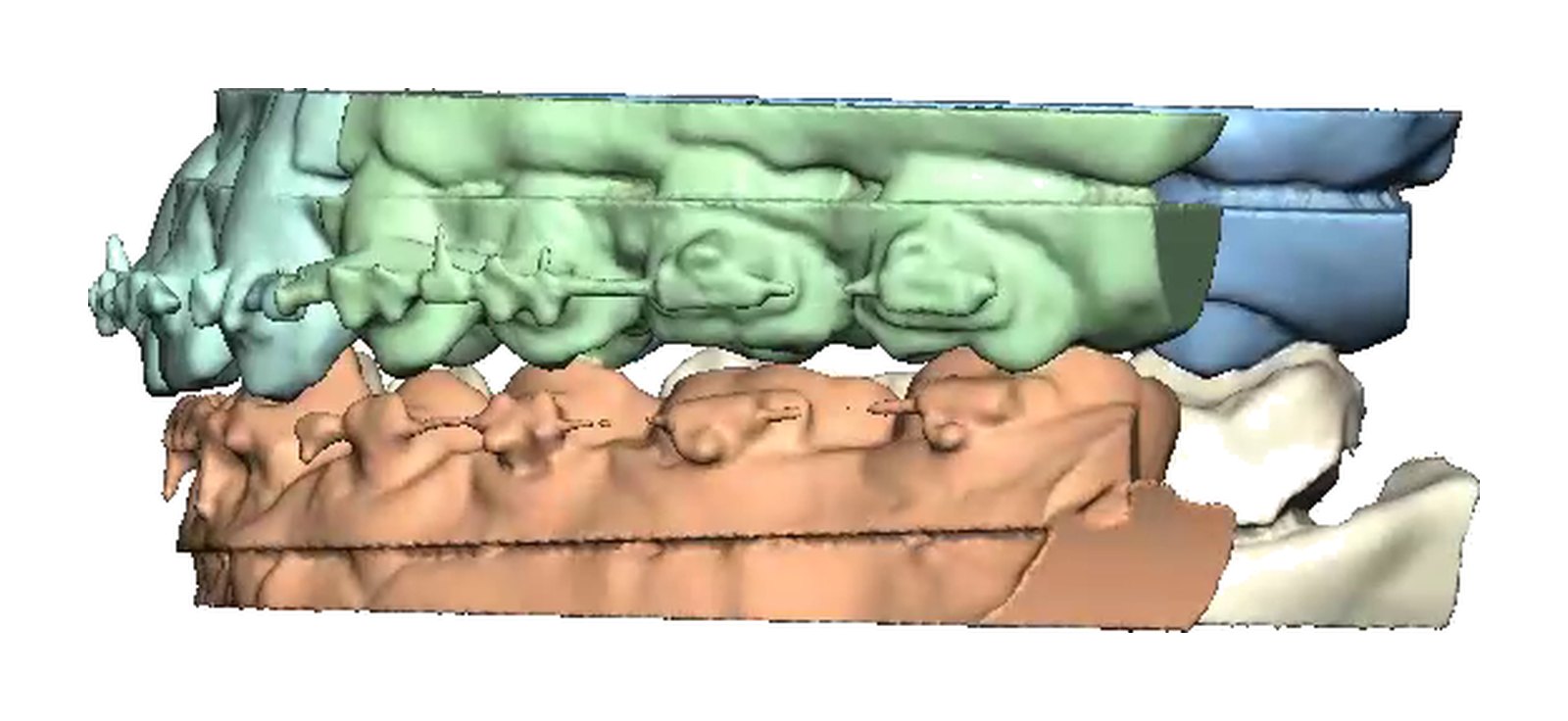
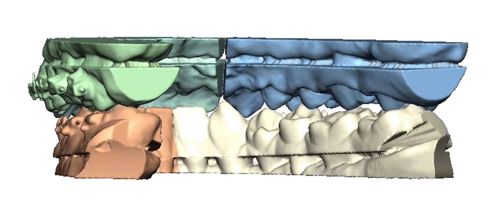
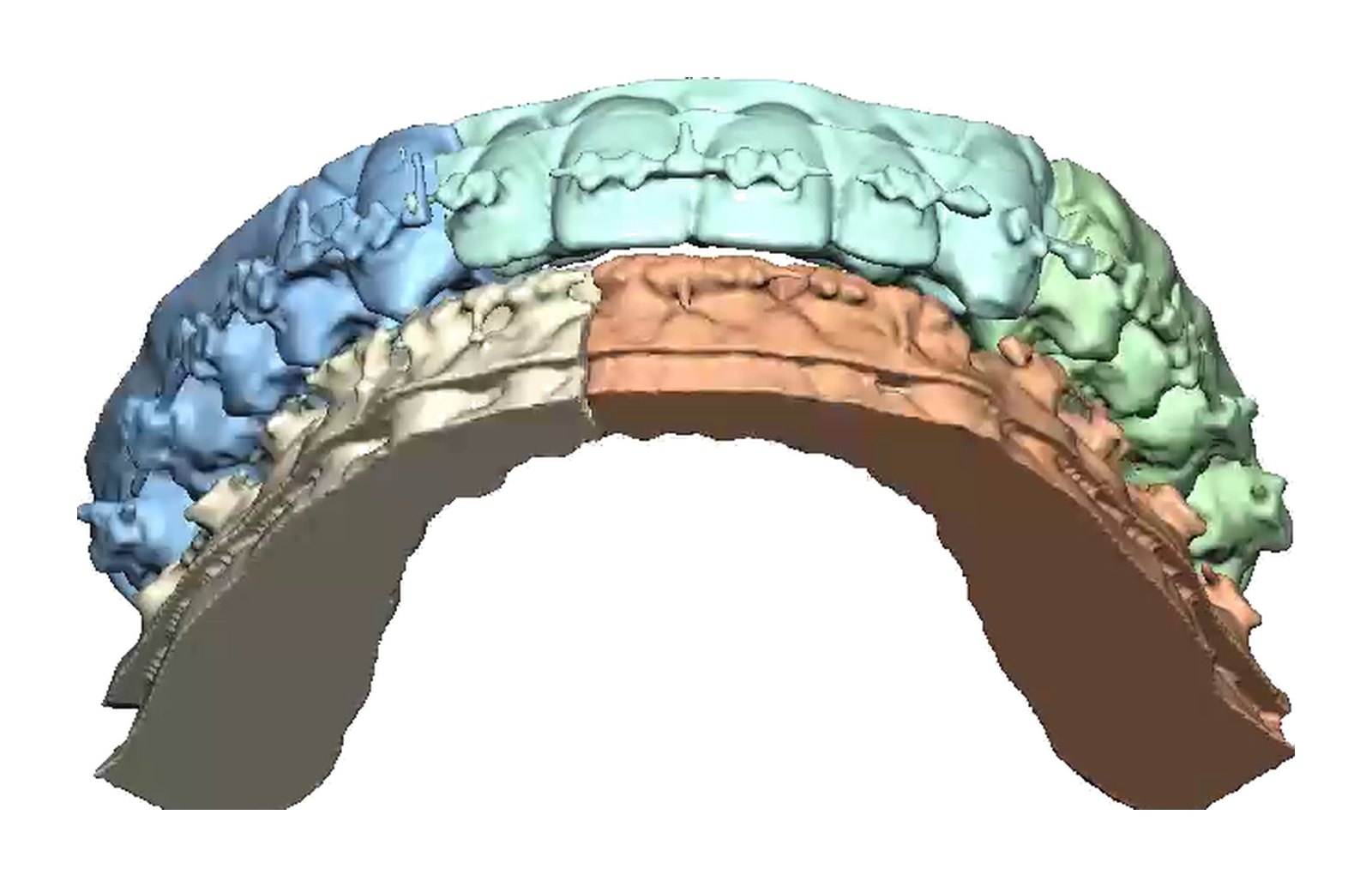
Planned final occlusion, frames from the 3D plan video:
Before & after (8 weeks)
Photographs before surgery and 8 weeks after surgery, at the same views. Orthodontic treatment was still in progress at 8 weeks.


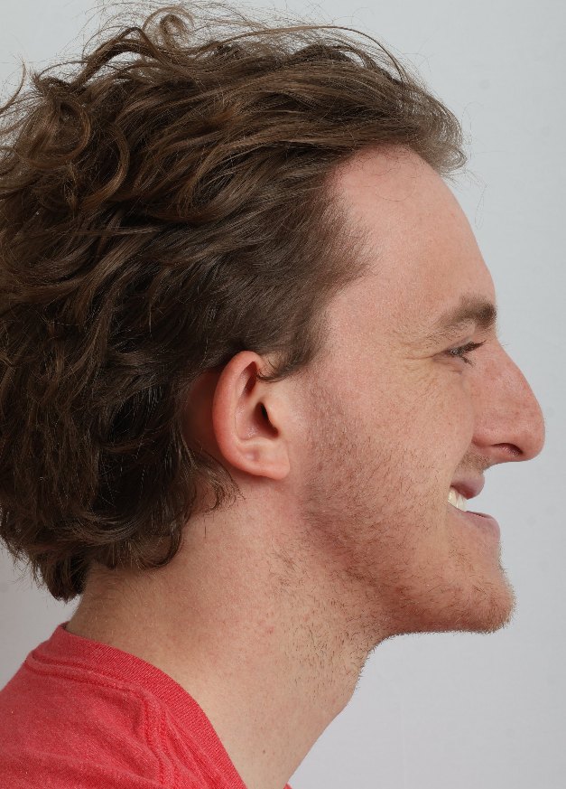
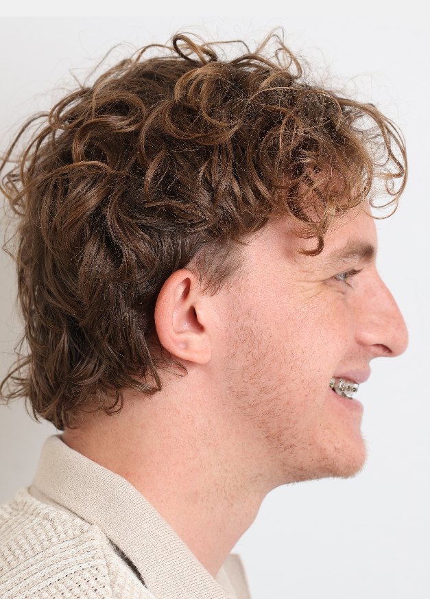



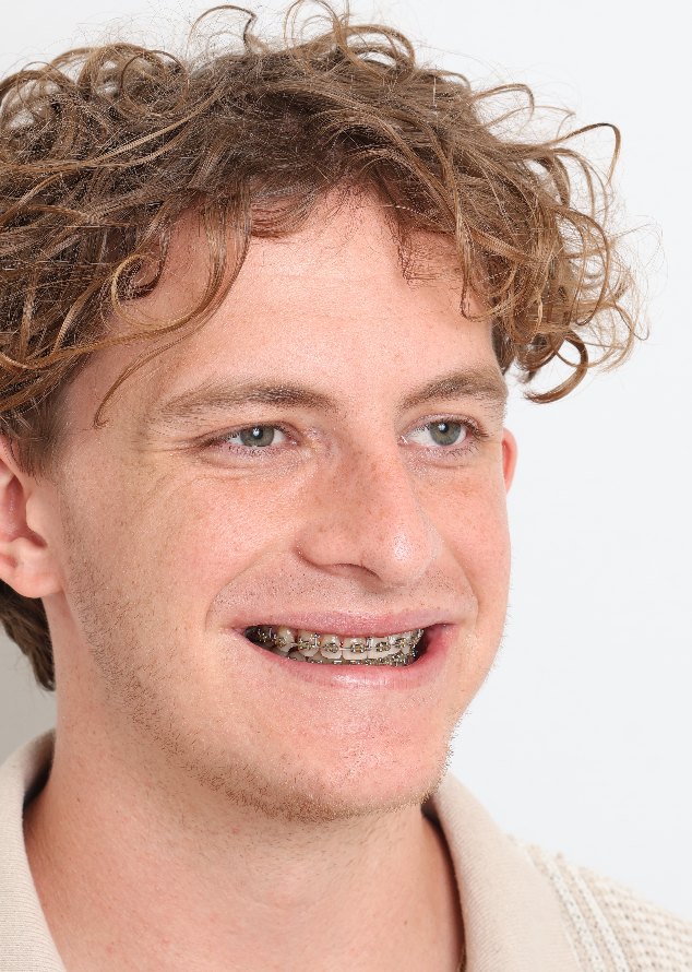
Early follow-up at 8 weeks; post-operative X-ray to be added. Individual results vary.
Where
Treated at Maxillo-Facial Surgery Institute, Irvine. Our Surgeons